

Dark circles under the eyes are one of the most common cosmetic concerns seen in clinical practice, yet they are frequently misunderstood. Medically referred to as periorbital hyperpigmentation or periorbital hyperchromia, the darkness that appears in the infraorbital region beneath the lower eyelid is rarely caused by a single factor. Understanding the underlying mechanism — whether vascular, pigmentary, structural, or a combination — is essential before choosing any treatment, because what works for one type may do little for another.
Key Takeaways
- Dark circles under the eyes have four recognised subtypes: vascular, pigmented, structural, and mixed — each with a distinct cause and treatment approach.
- Genetics, aging, sleep deprivation, allergies, sun exposure, and nutritional deficiencies are among the most clinically supported contributing factors.
- The colour of your dark circles — blue-purple, brown, or shadowed hollow — can help identify the likely underlying cause.
- Most dark circles are not a sign of serious illness, but persistent or sudden-onset darkness alongside other symptoms warrants a medical assessment.
- Effective management depends on correctly identifying the type; a qualified practitioner can use tools such as a Wood's lamp to assist diagnosis.
The Four Types of Dark Circles: A Starting Point for Understanding
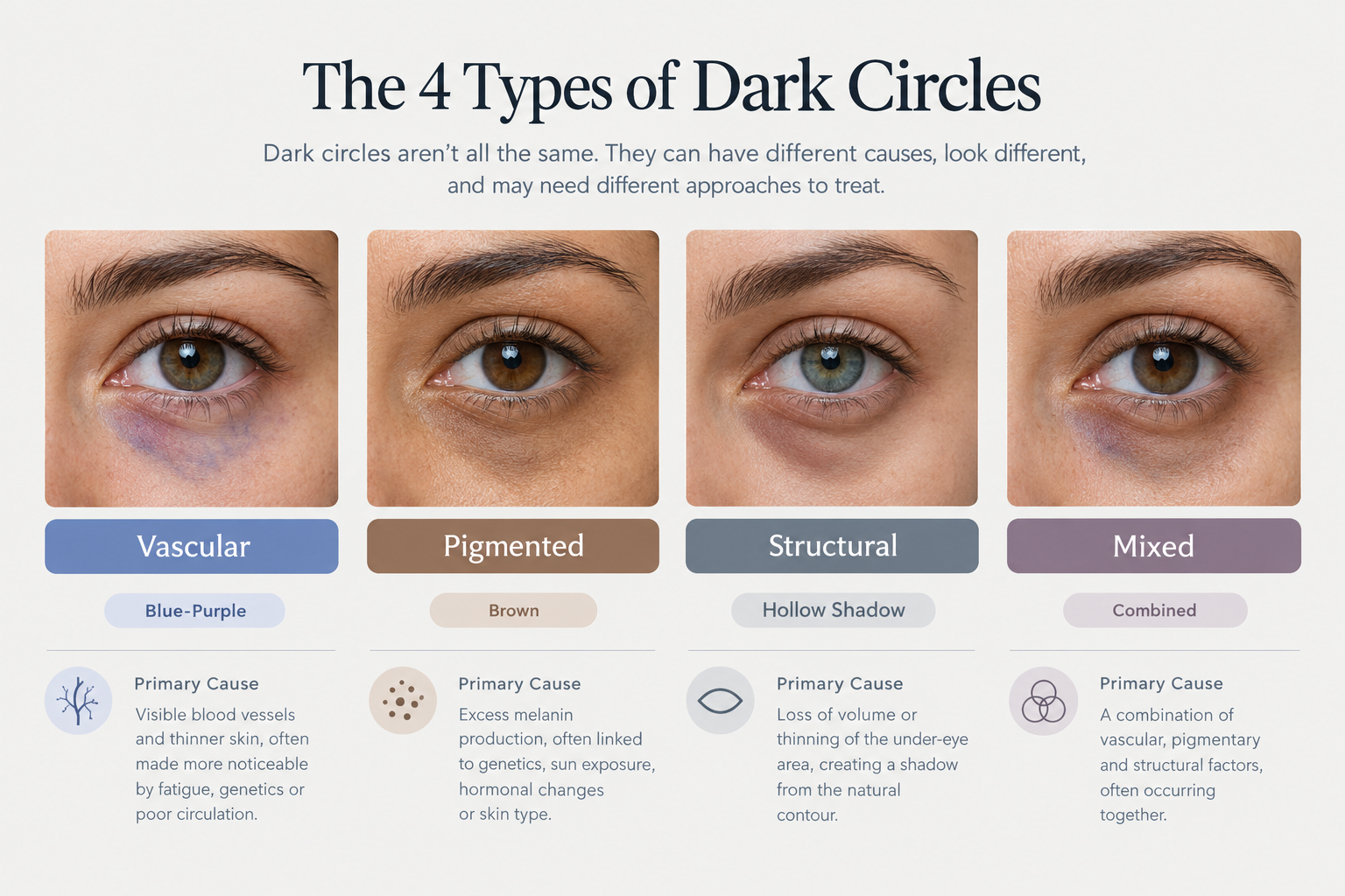
Clinicians generally classify periorbital darkness into four subtypes, and this classification matters because the cause — and therefore the most appropriate response — differs significantly between them. The three most widely recognised types are vascular, pigmented, and structural. A fourth category, mixed, is increasingly acknowledged in clinical literature and is arguably the most common presentation in practice, where two or more mechanisms overlap in the same patient.
Identifying your type is possible with a simple visual assessment. Blue or purple discolouration that appears worse in the morning typically suggests a vascular component. Brown or tan discolouration that is more uniform and does not change significantly with sleep points toward dermal melanin deposition. A hollow shadow beneath the eye that deepens with age or weight loss is characteristic of structural dark circles. When elements of more than one type are present simultaneously, the presentation is classified as mixed.
| Type | Typical Colour | Primary Cause | Common Treatment Direction |
|---|---|---|---|
| Vascular | Blue-purple | Blood stasis, visible vessels, poor microcirculation | Topical caffeine, vitamin K, laser (pulsed dye), lifestyle changes |
| Pigmented | Brown, tan | Dermal melanin deposition, post-inflammatory hyperpigmentation | Topical brighteners, chemical peels, Q-switched laser |
| Structural | Dark shadow | Volume loss, tear trough hollowing, skin laxity | Dermal fillers, PRP, blepharoplasty |
| Mixed | Variable | Combination of above mechanisms | Multimodal approach tailored to dominant component |
Vascular Causes: Blood Flow, Thin Skin, and Pooling
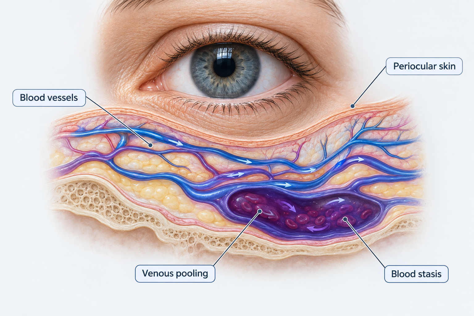
Vascular dark circles arise when blood and lymph microcirculation in the periocular skin becomes sluggish or impaired. The skin beneath the eye is among the thinnest on the entire body — often less than 0.5 mm — which means that underlying blood vessels are far more visible here than elsewhere on the face. When blood flow slows, pigments derived from haemoglobin breakdown, including bilirubin and other metabolites, accumulate in the tissue and produce the characteristic blue-violet hue seen in vascular dark circles.
Blood stasis and venous pooling are particularly pronounced during sleep. When lying flat, blood in the venous network beneath the eye pools due to reduced circulatory pressure, which is why vascular dark circles are often most noticeable immediately after waking. Sleep deprivation compounds this effect: fatigue causes blood vessels to dilate, increasing the volume of blood visible through the thin periorbital skin and making the area appear darker and more congested.
Nasal congestion and chronic sinus problems contribute to vascular dark circles through a related mechanism. The venous drainage from the periorbital region connects to the nasal and paranasal venous network. When this network becomes chronically congested — as occurs with persistent allergies or sinus disease — blood backs up into the vessels beneath the eye, producing the bluish discolouration commonly known as allergic shiners. This phenomenon is particularly common in children and adults with hay fever or perennial allergic rhinitis.
Certain prescription eye drops used to treat glaucoma, including bimatoprost and latanoprost, are a less commonly discussed but clinically recognised cause of periorbital darkening. These prostaglandin analogue medications can increase melanin production in the periocular skin and may also cause subtle changes in the surrounding tissue over time. Patients using these medications who notice progressive darkening around the eyes should raise this with their prescribing clinician.
Pigmented Causes: Melanin, Sun Exposure, and Skin Inflammation
Pigmented dark circles result from an excess of melanin — the pigment that gives skin its colour — deposited in the dermis or epidermis of the lower eyelid. This type of periorbital hyperpigmentation tends to appear brown or tan rather than blue, and it does not fluctuate significantly with sleep or hydration. It is more prevalent in individuals with naturally darker skin tones, partly because melanocytes (the cells that produce melanin) are more active in people with brown or Black skin, and partly because post-inflammatory hyperpigmentation (PIH) is more likely to leave lasting pigment changes in more pigmented skin.
Repeated UV exposure is one of the most consistent drivers of pigmented dark circles. Ultraviolet radiation stimulates melanocytes in the periorbital skin to produce more melanin as a protective response. Over time, this cumulative sun damage leads to uneven pigmentation that can be difficult to reverse without targeted treatment. Wearing broad-spectrum SPF around the eyes daily is one of the most evidence-supported preventive measures available.
Skin inflammation is another significant trigger for dermal melanin deposition beneath the eyes. Conditions such as atopic dermatitis (eczema), allergic contact dermatitis, and chronic eye rubbing all cause repeated low-grade trauma to the delicate periocular skin. Each episode of inflammation can leave behind post-inflammatory hyperpigmentation, which accumulates over months and years. This is why people who habitually rub their eyes — whether due to allergies, tiredness, or habit — often develop progressively darker under-eye pigmentation over time.
Hormonal factors may also influence pigmented dark circles, though this area is less extensively studied. Hormonal disturbances associated with pregnancy, oral contraceptive use, or thyroid dysfunction have been linked to worsening periorbital pigmentation in some individuals, possibly through effects on melanocyte-stimulating hormone activity. This overlap with melasma — a condition of hormonally driven facial hyperpigmentation — is worth noting, particularly in women who notice darkening that correlates with hormonal changes.
Structural Causes: Aging, Volume Loss, and the Tear Trough
Structural dark circles are caused not by pigment or blood vessels but by the three-dimensional anatomy of the face changing over time. As the face ages, collagen loss and reduced skin elasticity cause the skin beneath the eye to thin and loosen — a process known as skin laxity. Simultaneously, the fat pads that once provided smooth, full contour beneath the eye begin to diminish or shift, creating a concave hollow known as the tear trough. This hollow casts a shadow across the lower eyelid that mimics the appearance of dark pigmentation but is entirely structural in origin.
The tear trough deformity deepens with age and is also influenced by genetics and body weight. Individuals who lose significant weight may notice that structural dark circles worsen, as subcutaneous fat loss accelerates the hollowing effect. Conversely, periorbital oedema — puffiness or swelling of the eyelid — can create a contrasting shadow beneath the swollen area that also reads as darkness, even when no true pigmentation is present.
Collagen loss beneath the eye also allows blood vessels to become more visible through the increasingly translucent skin, which means structural and vascular components frequently coexist in older patients. This is one reason why the mixed subtype is so common in clinical practice: aging simultaneously thins the skin, reduces volume, and impairs microcirculation, producing a presentation that involves elements of all three primary types.
Genetics and Who Is Most Affected
A family history of dark circles is one of the strongest predictors of developing them. Genetic predisposition influences the thickness of periocular skin, the depth of the tear trough, the activity of melanocytes in the lower eyelid, and the efficiency of local microcirculation — all of which contribute to periorbital darkness. If both parents have prominent dark circles, their children are statistically more likely to develop them regardless of lifestyle factors.
Dark circles are more prevalent in elderly individuals, in people with darker skin tones, and in those with a family history of the condition. Children are not immune: because periocular skin is naturally thin and sensitive in early life, blood vessels beneath the eyes are often visible even in young children, particularly those with allergies or a tendency toward nasal congestion. In children, dark circles are most commonly vascular in origin and are frequently associated with allergic disease rather than lifestyle factors.
Ethnicity also influences the dominant subtype. Research suggests that in more darkly pigmented patients, the vascular component of dark circles may be underestimated because the brown skin tone can mask the blue-purple hue of venous pooling. Clinicians assessing patients with skin of colour need to consider that the visible discolouration may not accurately reflect the underlying mechanism, and that a predominantly vascular cause may be present even when the appearance looks pigmented.
Lifestyle and Environmental Factors
Several modifiable lifestyle factors contribute to dark circles, primarily through vascular and oxidative mechanisms. Sleep deprivation remains the most widely recognised lifestyle cause: insufficient sleep increases fluid retention around the eyes, dilates blood vessels, and reduces the skin's ability to recover from daily environmental stressors. While a single poor night's sleep rarely causes lasting change, chronic sleep deprivation can progressively worsen vascular dark circles over time.
Dehydration causes the skin to appear dull and sunken, making the periorbital area look darker and more hollow. Alcohol consumption worsens dehydration and also dilates blood vessels, compounding the vascular component. Chronic stress elevates cortisol levels, which can impair skin barrier function and microcirculation, contributing to a tired, darkened appearance around the eyes.
Environmental stressors — including UV radiation, cigarette smoke, and air pollution — generate oxidative stress in the skin through the production of free radicals. The delicate periocular skin is particularly susceptible to this oxidative damage, which can degrade collagen, impair microcirculation, and accelerate the visible signs of aging beneath the eyes. Smoking is a particularly significant contributor because it simultaneously causes oxidative stress, reduces skin oxygenation, and impairs collagen synthesis.
Nutritional Deficiencies and Dark Circles
Several nutritional deficiencies have been associated with periorbital darkness, though the strength of evidence varies between nutrients. Iron deficiency anaemia is the most clinically supported nutritional cause. Iron is essential for haemoglobin production, which carries oxygen to tissues throughout the body. When iron levels are insufficient, tissues receive less oxygen, small blood vessels dilate in an attempt to compensate, and the skin becomes paler overall — making the dark underlying vessels beneath the thin periorbital skin far more visible by contrast. A full blood count and iron studies can confirm whether iron deficiency is contributing.
Vitamin K plays a role in blood clotting and vascular integrity. When vitamin K levels are low, blood may be more prone to pooling in the small capillaries beneath the eyes rather than clearing efficiently, potentially worsening the vascular component of dark circles. Topical vitamin K is included in some under-eye formulations on this basis, though clinical evidence for topical application specifically remains limited.
Vitamins B12, C, and D have each been associated with periorbital darkness through different proposed mechanisms. Vitamin B12 deficiency can cause skin pallor that makes underlying vessels more visible, similar to iron deficiency. Vitamin C is essential for collagen synthesis; deficiency may accelerate skin thinning and reduce the structural support of periocular skin. Vitamin D's role is less clearly defined, and the evidence linking its deficiency specifically to dark circles is currently weaker than for iron or B12. If nutritional deficiency is suspected, a blood panel is the appropriate first step rather than supplementation without testing.
Medical and Systemic Conditions
While dark circles are most often a cosmetic rather than medical concern, certain systemic conditions can cause or worsen periorbital darkness and warrant clinical attention. Thyroid disease — both hypothyroidism and hyperthyroidism — can cause periorbital oedema and changes in skin texture that contribute to a darkened appearance. Chronic kidney disease may cause fluid retention and pallor that worsen the under-eye area. Anaemia from any cause, not only iron deficiency, can make dark circles more prominent by reducing skin oxygenation.
Allergic conditions deserve particular mention. Hay fever, perennial allergic rhinitis, and atopic dermatitis all contribute to dark circles through multiple pathways simultaneously: nasal congestion causes venous pooling (allergic shiners), chronic eye rubbing triggers post-inflammatory hyperpigmentation, and the inflammatory state of atopic skin can directly alter pigmentation in the periorbital region. Managing the underlying allergic condition often produces meaningful improvement in the appearance of dark circles without any cosmetic intervention.
How Dermatologists Diagnose the Type of Dark Circle
Accurate diagnosis of the dark circle subtype guides treatment selection and helps avoid ineffective or counterproductive interventions. A dermatologist or aesthetic practitioner will typically begin with a visual assessment under good lighting, examining the colour, distribution, and behaviour of the darkness (for example, whether it worsens with fatigue or remains constant). A simple stretch test — gently pulling the skin taut beneath the eye — can help distinguish pigmented from structural causes: if the darkness fades when the skin is stretched, it is more likely structural or vascular; if it persists, pigmentation is more likely.
A Wood's lamp — a UV light device used in dermatology — can help characterise the depth of pigmentation. Epidermal melanin fluoresces more intensely under Wood's lamp examination than dermal melanin, which helps the clinician determine whether pigment is superficial (and therefore more responsive to topical treatments) or deeper (requiring laser or other procedural approaches). Blood tests including a full blood count, ferritin, iron studies, and B12 levels may be requested if a nutritional deficiency is suspected as a contributing factor.
Treatment Directions: Matching the Approach to the Cause
Because the four subtypes of dark circles have distinct underlying mechanisms, treatment must be matched to the dominant cause. Applying a brightening serum to structural dark circles caused by tear trough hollowing, for example, is unlikely to produce meaningful improvement. Similarly, dermal filler placed in a predominantly pigmented dark circle may improve the shadow component but will not address the excess melanin.
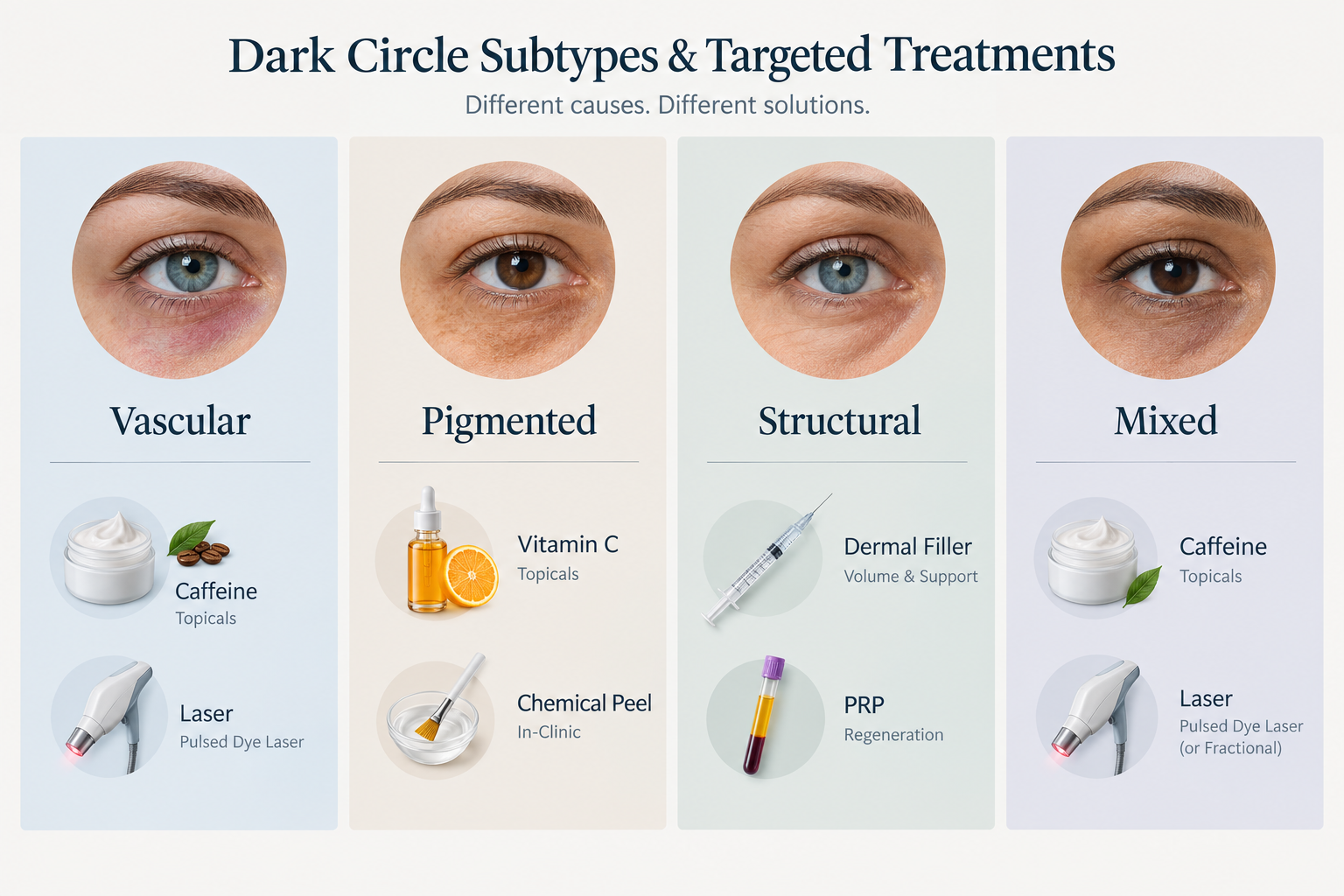
For vascular dark circles, topical ingredients such as caffeine (which constricts blood vessels), vitamin K, and niacinamide may offer modest improvement. Pulsed dye laser targets haemoglobin in superficial vessels and can reduce the blue-purple discolouration in suitable candidates. Lifestyle modifications — improving sleep, reducing alcohol, managing allergies — address the root cause and should accompany any topical or procedural treatment.
For pigmented dark circles, topical brightening agents including vitamin C, kojic acid, arbutin, tranexamic acid, niacinamide, and retinol are commonly used to reduce melanin production and accelerate cell turnover. Chemical peels using glycolic acid can improve superficial pigmentation. Q-switched and Nd:YAG lasers are used for deeper or more resistant pigmentation, though these require careful patient selection, particularly in individuals with darker skin tones where the risk of post-treatment hyperpigmentation is higher.
For structural dark circles, hyaluronic acid dermal fillers placed in the tear trough can restore lost volume and reduce the shadow effect. Platelet-rich plasma (PRP) injections may improve skin quality and stimulate collagen production in the periocular area. In cases of significant skin laxity or fat prolapse, surgical blepharoplasty may be the most effective long-term option. All procedural treatments carry risks and require assessment by a qualified practitioner to determine suitability.
Frequently Asked Questions
Can dark circles go away on their own?
Dark circles caused by temporary factors — such as a period of poor sleep, dehydration, or a short-term illness — often improve once the underlying cause resolves. However, dark circles driven by genetics, aging, structural volume loss, or chronic pigmentation are unlikely to resolve without targeted intervention. Identifying the cause is the most useful first step.
What deficiency most commonly causes dark circles?
Iron deficiency anaemia is the most clinically supported nutritional cause of dark circles. It causes pallor and vessel dilation that makes periorbital vessels more visible. Vitamin B12 deficiency can produce a similar effect. If deficiency is suspected, a blood test is the appropriate way to confirm this rather than self-supplementing without diagnosis.
Are dark circles hereditary?
Yes, genetics play a significant role. A family history of dark circles increases the likelihood of developing them, as inherited traits influence skin thickness, tear trough depth, melanocyte activity, and microcirculatory efficiency in the periorbital region. Hereditary dark circles may appear from childhood and tend to be persistent.
Do allergies cause dark circles?
Allergies are a well-recognised cause of vascular dark circles, particularly through the allergic shiner phenomenon. Chronic nasal congestion from hay fever or perennial allergic rhinitis causes venous pooling beneath the eyes. Additionally, allergic eye rubbing can trigger post-inflammatory hyperpigmentation over time. Treating the underlying allergy often improves the appearance of dark circles.
Can children get dark circles?
Yes. Children can develop dark circles for many of the same reasons as adults. Because periocular skin is naturally thin and sensitive in childhood, blood vessels beneath the eyes are often visible even in healthy children. In children, dark circles are most commonly associated with allergies, nasal congestion, or a genetic predisposition rather than lifestyle factors such as sleep deprivation or nutritional deficiency.
When should I see a doctor about dark circles?
Most dark circles are a cosmetic concern rather than a medical one. However, you should seek a medical assessment if dark circles appear suddenly without an obvious cause, if they are accompanied by significant swelling, pain, or vision changes, if you have other symptoms that suggest anaemia or thyroid disease, or if the darkness is asymmetric and worsening on one side only. A dermatologist can also assist with diagnosis and treatment planning for persistent dark circles that have not responded to lifestyle changes.
Understanding Your Dark Circles Before Seeking Treatment
Dark circles under the eyes are rarely caused by a single factor, and the most common mistake in managing them is treating the wrong type. A vascular dark circle will not respond to a brightening cream designed for pigmentation; a structural hollow will not improve with a topical serum alone. Taking time to identify the likely subtype — using colour, behaviour, and the stretch test as initial guides — significantly improves the chance of choosing an approach that produces a meaningful result.
For many people, addressing modifiable lifestyle factors such as sleep quality, hydration, allergy management, and sun protection will produce noticeable improvement without any clinical intervention. Where dark circles are driven by genetics, aging, or established pigmentation, a consultation with a dermatologist or qualified aesthetic practitioner is the most reliable route to an accurate diagnosis and a treatment plan matched to the actual cause. A Wood's lamp assessment and targeted blood tests can provide useful clinical information before any treatment is started, helping to avoid ineffective interventions and set realistic expectations.
