
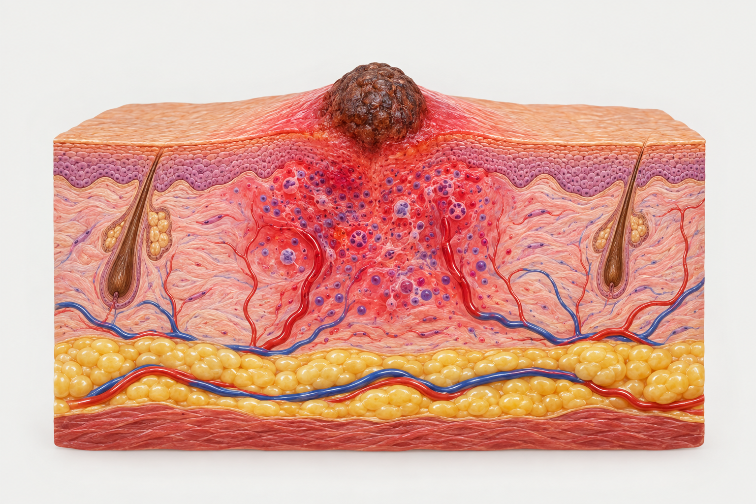

A mole that becomes red, swollen, painful, or begins to ooze can be alarming, and it is important to understand what these changes may mean. While some mole changes are caused by minor irritation or infection, others can signal something that requires prompt medical evaluation. This article explains the symptoms associated with an infected mole, what typically causes them, how infection differs from other concerning mole changes, and when professional assessment is essential.
Key Takeaways
- Redness, swelling, warmth, pain, and discharge around a mole are the most common signs of a localised skin infection near or involving a mole.
- An infected mole and a changing mole that may indicate melanoma or another skin cancer can look similar; professional assessment is always necessary to distinguish between them.
- Moles do not typically become infected on their own — infection usually follows trauma, scratching, shaving, or picking.
- Any mole that changes in colour, shape, size, or texture — with or without signs of infection — should be assessed by a qualified clinician without delay.
- Self-treatment of a suspected infected mole is not recommended; a clinician can determine whether the cause is infection, inflammation, or a more serious skin change.
What Does an Infected Mole Look Like?
An infected mole typically shows signs consistent with localised skin infection rather than changes to the mole itself. The surrounding skin may appear red and inflamed, and the area may feel warm or tender to the touch. Swelling around the base of the mole is common, and in some cases a small amount of clear, yellow, or green discharge may be present if a bacterial infection has developed. The mole may also feel sore when pressed or when clothing rubs against it.
It is worth noting that the mole itself — its pigmentation, borders, and structure — may not change significantly during a straightforward infection. The visible changes are usually in the skin immediately surrounding the mole rather than within the mole’s own tissue. However, because inflammation can temporarily alter the appearance of a mole, it can be difficult for a non-specialist to assess whether the underlying mole has also changed. This is one of the key reasons why a clinical examination is important whenever a mole appears infected.
In more advanced cases, a small abscess or crust may form at the site, and the area may become increasingly painful. Systemic symptoms such as fever, spreading redness, or swollen lymph nodes nearby would suggest the infection has extended beyond the immediate skin surface and requires urgent medical attention.
Common Causes of Mole Infection
Moles do not become infected spontaneously. Infection typically occurs when the skin barrier around or over a mole is disrupted, allowing bacteria — most commonly Staphylococcus aureus or Streptococcus species — to enter the tissue. The most frequent causes include accidental trauma, such as catching a raised mole on clothing or jewellery, shaving over a mole on the face, neck, or legs, and repeated scratching or picking at a mole that has become itchy.
Raised or pedunculated moles (those that sit above the skin surface on a stalk) are particularly vulnerable to trauma because they are more easily caught or rubbed. A mole that has been partially torn or cut is at higher risk of bacterial contamination. In some cases, a hair follicle within or adjacent to a mole may become blocked and infected, producing a folliculitis-like presentation that can be mistaken for mole infection.
People who have eczema or other inflammatory skin conditions may also experience irritation around moles due to scratching, which can secondarily introduce bacteria. Immunocompromised individuals may be at greater risk of skin infections generally, including around moles.
Infected Mole vs Skin Cancer: Understanding the Difference
One of the most important clinical distinctions is between an infected mole and a mole that is changing due to a potentially malignant process such as melanoma. Both can cause visible changes to a mole’s appearance, and some features can overlap, which is why self-diagnosis is unreliable and professional evaluation is essential.
Melanoma and other skin cancers typically cause changes within the mole itself rather than in the surrounding skin. Clinicians commonly use the ABCDE criteria as a framework for identifying mole changes that warrant further investigation:
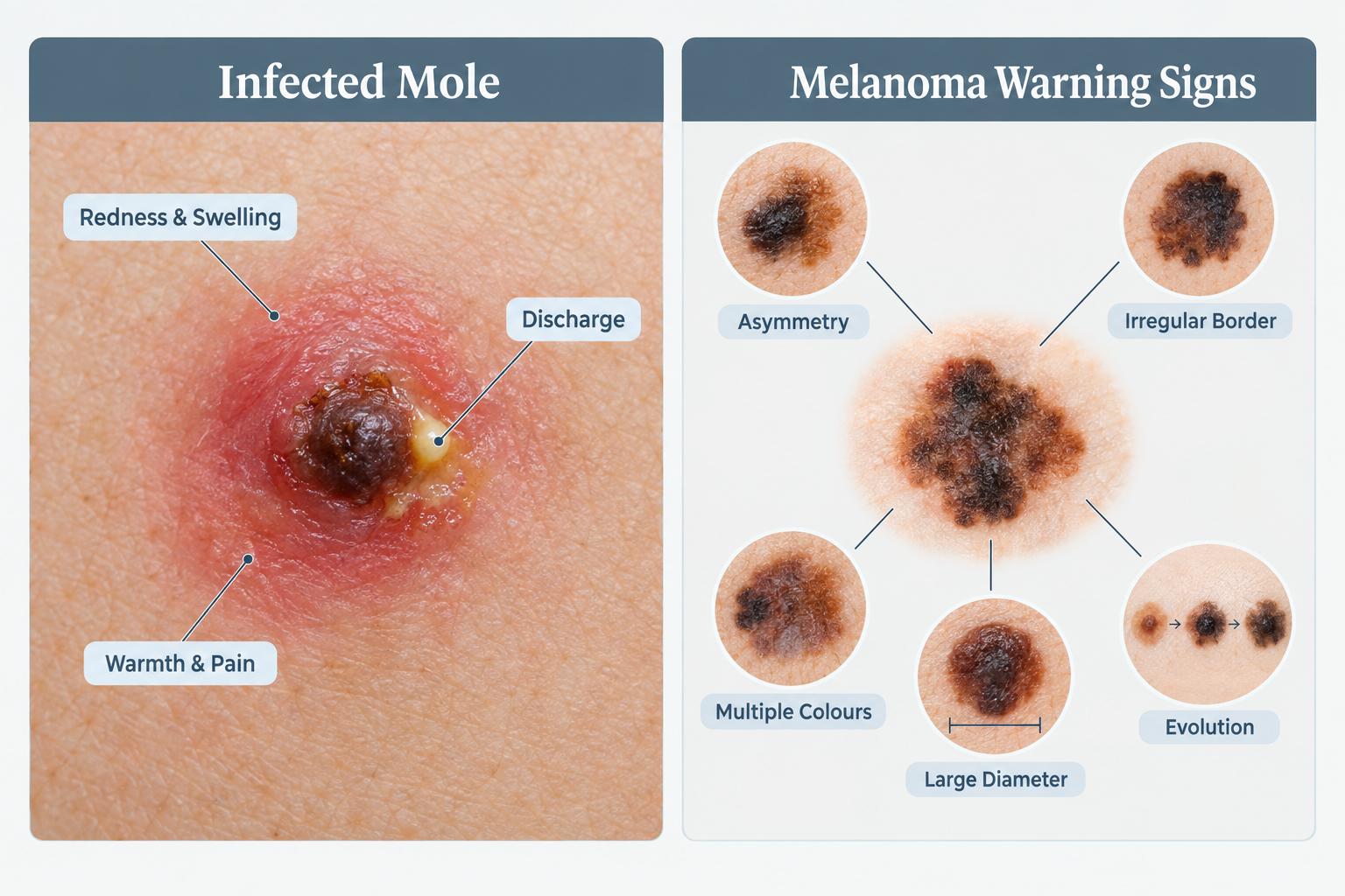
- Asymmetry: One half of the mole does not match the other.
- Border: The edges are irregular, ragged, notched, or blurred.
- Colour: The mole contains multiple shades of brown, black, red, white, or blue.
- Diameter: The mole is larger than approximately 6 mm, though melanomas can be smaller.
- Evolution: The mole is changing in size, shape, colour, or any other characteristic, including new symptoms such as bleeding or itching.
An infected mole, by contrast, tends to show signs of acute inflammation in the surrounding skin — redness, warmth, swelling, and discharge — rather than structural changes within the mole’s pigmented tissue. However, inflammation can temporarily distort a mole’s appearance, making it harder to assess the underlying structure. A clinician may recommend treating the infection first and then re-examining the mole once the inflammation has resolved, or may refer the patient for dermoscopy or biopsy if there is any clinical uncertainty.
It is also important to be aware that amelanotic melanoma — a form of melanoma that lacks the typical dark pigmentation — can sometimes resemble an inflamed or infected skin lesion. This further underlines why any mole that is changing, bleeding, or causing new symptoms should be assessed by a qualified clinician rather than assumed to be a simple infection.
Symptoms That Require Urgent Assessment
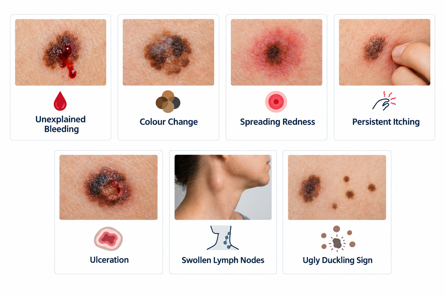
Certain symptoms associated with a mole change should prompt prompt medical review rather than a wait-and-see approach. These include:
- A mole that bleeds without obvious trauma or that bleeds repeatedly.
- A mole that has changed noticeably in size, shape, or colour over a period of weeks to months.
- A mole that has become persistently itchy, crusted, or ulcerated.
- Signs of spreading infection, including expanding redness (cellulitis), fever, or swollen lymph nodes in the neck, armpit, or groin near the affected mole.
- A new dark streak or pigmentation extending from the mole into the surrounding skin.
- A mole that has developed a raised, firm, or nodular area within it.
- Any mole that looks significantly different from your other moles — sometimes called the “ugly duckling” sign.
These features do not automatically confirm a serious diagnosis, but they do indicate that a clinical assessment — and potentially dermoscopy or a skin biopsy — is necessary. Early assessment of changing moles is associated with better outcomes when skin cancer is identified, which is why prompt action is always advisable.
How a Clinician Assesses a Suspected Infected Mole
When a patient presents with a mole that appears infected or has changed, a clinician will typically begin with a detailed history. This includes how long the changes have been present, whether there was any preceding trauma, whether the mole has changed over time, and whether the patient has a personal or family history of skin cancer or atypical moles.
A physical examination of the mole and surrounding skin will follow. Dermoscopy — a non-invasive technique using a handheld magnifying device with polarised light — allows a clinician to examine the internal structures of a mole in greater detail than is possible with the naked eye. Dermoscopy can help distinguish between inflammatory changes and structural features associated with malignancy, though it requires specialist training to interpret accurately.
If infection is confirmed and the mole’s underlying structure appears benign, treatment for the infection will typically be recommended. If there is any clinical uncertainty about the mole itself, the clinician may arrange a skin biopsy. A biopsy involves removing a small sample of tissue — or in some cases the entire mole — for laboratory analysis. This is the definitive method for determining whether a mole contains abnormal or malignant cells.
In some cases, a clinician may refer the patient to a dermatologist or a specialist skin cancer clinic for further evaluation, particularly if the mole has features that are difficult to assess in a primary care setting.
Treatment Options for an Infected Mole
The appropriate treatment for an infected mole depends on the severity of the infection and whether the mole itself requires further investigation. For a mild localised infection, a clinician may recommend a course of topical or oral antibiotics to clear the bacterial infection. Keeping the area clean and avoiding further trauma — such as shaving over the mole or wearing tight clothing that rubs the site — is also important during recovery.
If an abscess has formed, a clinician may need to drain it. This is a minor procedure performed under local anaesthetic and should not be attempted at home, as improper drainage can worsen infection or introduce additional bacteria.
Once the infection has resolved, the mole should be re-examined to assess its underlying appearance. If the mole was already being monitored for atypical features, or if the clinician has any concerns following re-examination, removal of the mole may be recommended. Mole removal is typically performed by a dermatologist or trained clinician using surgical excision, shave excision, or another appropriate technique depending on the mole’s characteristics. The removed tissue is usually sent for histological analysis to confirm the diagnosis.
It is important to understand that over-the-counter creams, home remedies, or attempts to remove a mole at home are not appropriate responses to a suspected infected or changing mole. These approaches can delay proper diagnosis, cause scarring, and — in the case of a mole that turns out to be malignant — allow a serious condition to progress without treatment.
Can You Prevent Mole Infection?
While it is not always possible to prevent a mole from becoming infected, certain practical steps can reduce the risk of trauma and subsequent infection, particularly for raised or prominent moles.
- Avoid shaving directly over raised moles; use an electric razor or shave around the mole carefully.
- Protect moles from repeated friction by choosing clothing and accessories that do not rub against them.
- Avoid picking, scratching, or attempting to remove a mole yourself.
- Keep the skin around moles clean and moisturised, particularly if you have a skin condition such as eczema that increases the risk of scratching.
- If a mole is accidentally cut or torn, clean the area gently with mild soap and water, apply a clean dressing, and seek medical advice if signs of infection develop.
Regular skin self-examination is also a useful habit. Becoming familiar with your moles — their usual size, shape, colour, and texture — makes it easier to notice when something has changed. Many dermatology organisations recommend checking your skin monthly and having a professional skin examination at intervals recommended by your clinician, particularly if you have a large number of moles, a history of sun damage, or a family history of melanoma.
The Role of Sun Protection in Mole Health
Sun exposure is the most significant modifiable risk factor for melanoma and other skin cancers. Ultraviolet radiation from the sun and from artificial sources such as sunbeds can cause DNA damage in skin cells, increasing the risk that a mole or area of normal skin will develop malignant changes over time. While sun exposure does not directly cause mole infection, protecting moles from UV damage is an important part of overall skin health and cancer prevention.
Broad-spectrum sunscreen with an SPF of 30 or higher, applied consistently to exposed skin including areas with moles, is recommended by dermatology guidelines. Protective clothing, hats, and avoiding peak UV hours also contribute to reducing cumulative sun damage. People with a large number of moles, fair skin, or a personal or family history of skin cancer may benefit from discussing a personalised sun protection and monitoring plan with a dermatologist.
Frequently Asked Questions
Can a mole become infected without any obvious injury?
It is uncommon for a mole to become infected without some form of skin disruption, even if the cause is not immediately obvious. Minor trauma such as unconscious scratching during sleep, friction from clothing, or a small insect bite near the mole can be enough to break the skin barrier and allow bacteria to enter. If a mole appears infected without a clear cause, a clinician should assess it to rule out other explanations for the changes.
How long does it take for an infected mole to heal?
With appropriate antibiotic treatment and care, a mild mole infection typically begins to improve within a few days and resolves within one to two weeks. More significant infections, or those involving an abscess, may take longer and may require additional treatment. If the area is not improving with treatment, or if symptoms are worsening, further medical review is necessary.
Should I be worried if my mole is itchy but not visibly infected?
Persistent itching in a mole — particularly if it is a new symptom — is one of the features included in clinical assessments of changing moles and should be evaluated by a clinician. Itching alone does not confirm infection or malignancy, but it is a symptom that warrants professional assessment rather than self-monitoring at home.
Is it safe to have a mole removed if it has been infected?
Clinicians generally prefer to treat an active infection before performing elective mole removal, as operating on infected tissue carries a higher risk of complications including poor wound healing and spreading infection. Once the infection has resolved and the skin has healed, mole removal can be considered if clinically indicated. Your clinician will advise on the appropriate timing based on your individual situation.
What is dermoscopy and why might it be used for a mole?
Dermoscopy is a non-invasive diagnostic technique in which a clinician uses a handheld device with magnification and polarised light to examine the internal structures of a mole or skin lesion. It allows more detailed assessment of features such as pigment patterns, vascular structures, and border characteristics that are not visible to the naked eye. Dermoscopy is widely used in dermatology to improve the accuracy of mole assessment and to help distinguish benign moles from those that may require biopsy.
Can children’s moles become infected?
Yes, moles in children can become infected in the same way as those in adults, typically following trauma such as scratching or catching a raised mole. Any mole in a child that shows signs of infection, or that has changed in appearance, should be assessed by a clinician. Congenital moles — those present from birth — may be larger and are sometimes monitored more closely; any changes to these should be reported to a clinician promptly.
When to Act: Making the Right Decision About a Changing Mole
A mole that shows signs of infection — redness, swelling, warmth, pain, or discharge — should be assessed by a clinician rather than managed at home. While many cases turn out to involve straightforward bacterial infection following minor trauma, the clinical overlap between an infected mole and a mole undergoing malignant change means that self-diagnosis is not reliable. A qualified clinician can examine the mole, determine the most likely cause, arrange appropriate treatment, and decide whether further investigation such as dermoscopy or biopsy is needed.
If you notice any of the ABCDE warning signs — asymmetry, irregular borders, multiple colours, increasing diameter, or evolution of any kind — alongside or independently of signs of infection, seek medical assessment promptly. Early identification of skin cancer, including melanoma, is associated with significantly better treatment outcomes. Equally, if a mole is bleeding, persistently itching, crusting, or simply looks different from how it has always appeared, these are valid reasons to seek a professional opinion without delay.
Routine skin monitoring, consistent sun protection, and awareness of your own moles are practical steps that support long-term skin health. If you have concerns about a mole — infected or otherwise — a consultation with a dermatologist or trained clinician is the appropriate first step.
