


Acrochordons, more commonly known as skin tags, are among the most frequently encountered benign skin growths in adults. Although they are harmless in the vast majority of cases, they can cause discomfort when they catch on clothing or jewellery, and many people seek removal for cosmetic reasons. This article explains what acrochordons are, why they develop, how a qualified practitioner may assess and remove them, and what realistic outcomes look like.
Key Takeaways
- Acrochordons are benign, soft, pedunculated skin growths that pose no medical risk in the overwhelming majority of cases, but professional assessment is advisable before any removal attempt.
- They are strongly associated with friction, insulin resistance, obesity, and hormonal changes, meaning recurrence is possible if underlying factors are not addressed.
- Several effective removal methods exist, including cryotherapy, electrocautery, snip excision, and laser treatment; the most appropriate option depends on lesion size, location, and individual health factors.
- Home removal attempts carry real risks including bleeding, infection, and scarring, and are not recommended by dermatologists.
- Any skin lesion that changes in colour, bleeds spontaneously, or is uncertain in diagnosis should be assessed by a qualified clinician before treatment.
What Is an Acrochordon?
An acrochordon is a small, soft, flesh-coloured or slightly pigmented outgrowth of skin attached to the body by a narrow stalk called a peduncle. The term comes from the Greek words for extremity and cord, which describes the characteristic hanging appearance of these lesions. In clinical settings they may also be referred to as fibroepithelial polyps, soft fibromas, or cutaneous papillomas, though skin tag remains the most widely understood term among patients.
Histologically, an acrochordon consists of a loose fibrovascular core covered by a thin layer of epidermis. The stalk contains collagen fibres and small blood vessels, which is why larger tags can bleed if traumatised. Most acrochordons are between one and five millimetres in diameter, though some larger variants, sometimes called giant acrochordons, can reach a centimetre or more. They are almost always benign and do not carry a risk of malignant transformation, but this does not mean every suspicious lesion should be assumed to be a skin tag without professional evaluation.
Where Do Skin Tags Typically Appear?
Acrochordons develop most commonly in areas where skin rubs against skin or clothing. The neck, armpits, groin, eyelids, and the skin beneath the breasts are the most frequently affected sites. The inframammary fold and inner thighs are also common locations, particularly in individuals with higher body weight. Skin tags on the eyelids, known as periorbital acrochordons, require particular care during removal because of the proximity to the eye and the delicacy of the surrounding tissue.
Multiple acrochordons are common; it is not unusual for a person to develop dozens over time, particularly if predisposing factors such as obesity or insulin resistance are present. The distribution tends to follow friction zones, which supports the mechanical irritation theory of their development.
Why Do Acrochordons Form?
The precise mechanism behind acrochordon formation is not fully established, but several contributing factors are well recognised in the dermatological literature. Chronic skin-to-skin or skin-to-clothing friction is considered a primary driver, which explains why these lesions cluster in body folds and areas of repeated movement.
Insulin resistance and type 2 diabetes are strongly associated with multiple acrochordons. Research has found that the presence of numerous skin tags may serve as a clinical marker for impaired glucose metabolism, and some clinicians consider a sudden increase in skin tag number a reason to screen for metabolic conditions. The proposed mechanism involves elevated insulin and insulin-like growth factor levels stimulating epidermal and dermal proliferation.
Hormonal changes during pregnancy can also trigger the development of new acrochordons, and they may partially resolve after delivery. Obesity increases both friction and the hormonal environment that promotes soft tissue proliferation. There is also evidence of a familial tendency, suggesting a genetic predisposition in some individuals. Human papillomavirus (HPV) has been detected in some acrochordon tissue samples, though its causal role remains debated and acrochordons are not considered contagious in the way that viral warts are.
Are Skin Tags Dangerous?
The vast majority of acrochordons are entirely benign and require no treatment from a purely medical standpoint. They do not become cancerous, and they do not spread to other areas of the body. However, there are important reasons to seek professional assessment rather than assuming any skin lesion is a harmless skin tag.
Certain skin conditions can resemble acrochordons but have different clinical significance. Seborrhoeic keratoses, dermatofibromas, neurofibromas, and in rare cases early melanoma or other malignant lesions can superficially resemble a skin tag. A qualified clinician, typically a dermatologist or experienced aesthetic practitioner, can use dermoscopy and clinical examination to distinguish between these possibilities. Any lesion that bleeds without trauma, changes colour or shape rapidly, becomes ulcerated, or is associated with pain should be assessed promptly rather than treated as a routine skin tag.
Acrochordons can become uncomfortable or painful if they undergo torsion, meaning the stalk twists and cuts off the blood supply. This can cause the tag to become dark, swollen, and tender before it eventually falls off. While this process is not dangerous, it can be distressing and may warrant professional management.
Diagnosis and Clinical Assessment
Diagnosis of an acrochordon is usually straightforward through visual inspection and dermoscopy. A practitioner will examine the lesion’s surface texture, colour, attachment point, and surrounding skin. Dermoscopy, which uses a handheld magnifying device with polarised light, allows closer inspection of the lesion’s internal structure and helps rule out pigmented or vascular lesions that might otherwise be mistaken for a skin tag.
In most cases, histological analysis of removed tissue is not required for a typical small acrochordon. However, if a lesion has atypical features, is unusually large, or the clinician has any diagnostic uncertainty, the removed tissue may be sent for pathological examination. This is a routine precaution rather than a cause for alarm.
Acrochordon Removal: Available Treatment Options
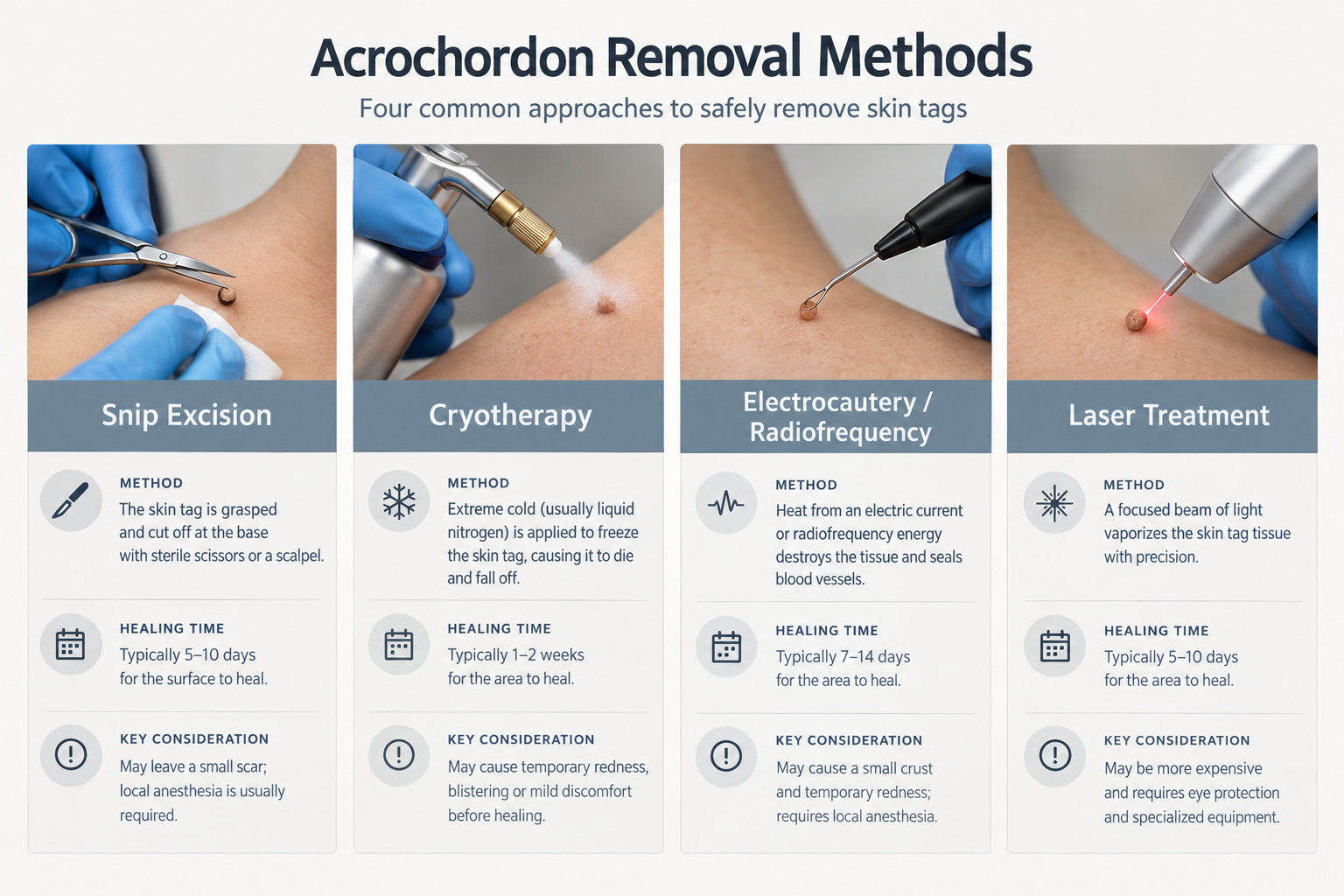
When removal is desired, whether for comfort or cosmetic reasons, several clinically established methods are available. The most appropriate technique depends on the size and location of the acrochordon, the number of lesions, the patient’s skin type, and any relevant medical history. A qualified practitioner will assess these factors before recommending a specific approach.
Snip Excision
Snip excision involves using sterile surgical scissors or a scalpel to cut the acrochordon at its stalk. This is one of the most commonly used methods for small to medium-sized skin tags and can be performed quickly in a clinical setting, often with or without a small amount of local anaesthetic depending on the size and sensitivity of the area. The procedure is precise, allows the removed tissue to be sent for histology if needed, and typically results in minimal scarring when performed by an experienced practitioner. Minor bleeding at the site is common and is usually controlled with pressure or a haemostatic agent.
Cryotherapy
Cryotherapy uses liquid nitrogen to freeze the acrochordon, causing cellular destruction and eventual shedding of the lesion over one to two weeks. It is a well-established dermatological technique suitable for small to medium acrochordons. The treated area may blister, crust, and temporarily darken or lighten depending on the patient’s skin tone. Post-inflammatory hyperpigmentation or hypopigmentation is a recognised risk, particularly in individuals with darker skin phototypes, and this should be discussed during the consultation. More than one session may be required for larger lesions.
Electrocautery and Radiofrequency
Electrocautery uses an electrical current to generate heat that destroys the acrochordon tissue and simultaneously seals the small blood vessels at the base. Radiofrequency devices work on a similar principle but use high-frequency alternating current, which some practitioners prefer for its precision and reduced thermal spread to surrounding tissue. Both methods are effective for a range of acrochordon sizes and can treat multiple lesions in a single session. Local anaesthetic is typically applied beforehand. Temporary redness, crusting, and mild swelling are expected in the days following treatment.
Laser Treatment
Laser removal of acrochordons uses targeted light energy to ablate or coagulate the lesion. Carbon dioxide (CO2) lasers and erbium lasers are among those used in clinical practice for this purpose. Laser treatment offers precision and is particularly useful for lesions in sensitive areas such as the eyelids or face. It may also be preferred when treating multiple lesions in a single session. As with other methods, temporary redness, crusting, and pigmentary changes are possible side effects. Laser treatment for skin tags is generally considered a cosmetic procedure and is not typically covered by health insurance.
Ligation
Ligation involves tying a fine suture or thread tightly around the base of the acrochordon stalk, cutting off its blood supply so that the lesion gradually necrotises and falls away over several days. This technique is sometimes used in clinical settings for pedunculated lesions with a clearly defined stalk. It is not recommended as a home remedy because of the risk of infection, incomplete removal, and the difficulty of correctly identifying the lesion without professional assessment.
What Happens During a Removal Appointment?
A typical acrochordon removal appointment begins with a clinical assessment of the lesion or lesions to confirm the diagnosis and discuss the most appropriate removal method. The practitioner will review relevant medical history, including any blood-thinning medications, bleeding disorders, or skin conditions that might affect healing. Photographs may be taken for clinical records.
For most small acrochordons, the procedure itself takes only a few minutes per lesion. Local anaesthetic cream or an injectable local anaesthetic may be applied depending on the method and the sensitivity of the area. Most patients describe the procedure as mildly uncomfortable rather than painful. After removal, the site is cleaned and a small dressing may be applied. Patients are usually able to return to normal activities immediately or within a day, depending on the location and extent of treatment.
Recovery and Aftercare
Recovery after acrochordon removal is generally straightforward. The treated area typically develops a small scab or crust that heals within one to two weeks. Keeping the area clean and dry, avoiding picking at the crust, and applying any recommended topical preparation as directed by the practitioner supports healing and reduces the risk of infection or scarring.
Sun protection is important for treated areas, particularly on the face and neck, as UV exposure to healing skin can worsen post-inflammatory pigmentation. A broad-spectrum sunscreen should be applied once the skin has re-epithelialised. Strenuous exercise or activities that cause significant sweating in the treated area may be temporarily restricted, particularly if the removal site is in a skin fold. The practitioner will provide specific aftercare guidance based on the method used and the location of the treated lesions.
Risks and Potential Side Effects
Acrochordon removal is generally considered a low-risk procedure when performed by a qualified clinician, but no procedure is entirely without risk. Patients should be aware of the following possibilities before proceeding.
- Scarring: Any skin procedure carries a small risk of scarring. The risk is generally low for small acrochordons removed by an experienced practitioner, but individuals with a history of keloid or hypertrophic scarring should discuss this with their clinician before treatment.
- Pigmentary changes: Temporary or, less commonly, longer-lasting changes in skin colour at the treatment site are possible, particularly with cryotherapy or laser. Darker skin phototypes carry a higher risk of post-inflammatory hyperpigmentation.
- Infection: Any break in the skin carries a small risk of infection. Signs of infection include increasing redness, warmth, swelling, discharge, or fever, and should be reported to the treating practitioner promptly.
- Incomplete removal: Occasionally a lesion may not be fully removed in a single treatment, particularly with cryotherapy, and a repeat session may be needed.
- Recurrence: Removing an acrochordon does not prevent new ones from forming, particularly if underlying factors such as friction, obesity, or insulin resistance remain present.
- Bleeding: Minor bleeding at the removal site is common and usually self-limiting. Larger acrochordons with a more substantial blood supply may bleed more, and the practitioner will take appropriate steps to manage this during the procedure.
Why Home Removal Is Not Recommended
A significant number of people attempt to remove acrochordons at home using thread, nail clippers, or over-the-counter freezing kits. Dermatologists and aesthetic practitioners consistently advise against this approach for several reasons. First, without professional assessment there is no certainty that the lesion is a benign acrochordon rather than a different type of skin growth that requires different management. Second, home removal attempts frequently result in incomplete removal, infection, excessive bleeding, and scarring. Third, over-the-counter cryotherapy products designed for wart removal are not formulated or calibrated for acrochordon removal and may cause tissue damage to the surrounding skin.
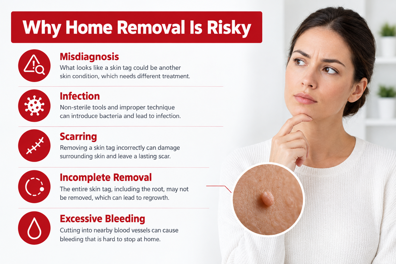
Professional removal in a clinical setting provides accurate diagnosis, appropriate technique selection, sterile conditions, and aftercare guidance that home methods cannot replicate. The cost of professional treatment is modest relative to the risks of complications from unsupervised attempts.
Who May Be Suitable for Acrochordon Removal?
Most adults with confirmed acrochordons who are bothered by their appearance or experiencing discomfort from friction or snagging are potential candidates for professional removal. Suitability for a specific removal method will depend on individual factors including the size, number, and location of lesions, skin type, medical history, and any medications that affect bleeding or healing.
Individuals taking anticoagulant medications should inform their practitioner, as this may influence the choice of technique and the management of any bleeding. Those with a history of keloid scarring, active skin infection in the treatment area, or certain autoimmune skin conditions may require a modified approach or may need to defer treatment. Pregnant individuals should discuss timing with their clinician, as some acrochordons that develop during pregnancy may partially resolve after delivery, and elective procedures are generally deferred where possible during pregnancy.
Alternatives to Professional Removal
For individuals who choose not to pursue professional removal, the main alternative is to leave the acrochordon in place and monitor it. Since acrochordons are benign, watchful waiting is a clinically reasonable option when the lesion is not causing discomfort or cosmetic concern. Addressing underlying contributing factors, such as achieving a healthier body weight, managing blood glucose levels, or reducing friction through appropriate clothing choices, may slow the development of new lesions, though it is unlikely to cause existing ones to disappear.
Some topical preparations marketed for skin tag removal are available without prescription, but the clinical evidence supporting their efficacy and safety is limited. These products should not be used without first confirming the diagnosis with a qualified clinician, and they are not a substitute for professional assessment of any lesion with atypical features.
Frequently Asked Questions
Do acrochordons grow back after removal?
Removing an acrochordon eliminates that specific lesion, but it does not prevent new ones from forming elsewhere. Individuals with predisposing factors such as obesity, insulin resistance, or significant skin friction are more likely to develop further acrochordons over time. Addressing these underlying factors where possible may help reduce the rate of new lesion development.
Is acrochordon removal painful?
Most removal procedures involve minimal discomfort when local anaesthetic is used. Without anaesthetic, snipping a small acrochordon may cause a brief sharp sensation. Cryotherapy can cause a stinging or burning feeling during and shortly after application. Most patients find the procedures tolerable, and any discomfort is typically short-lived.
How long does healing take after skin tag removal?
Most treated sites heal within one to two weeks, forming a small scab that gradually falls away to reveal new skin beneath. Pigmentary changes, if they occur, may take several weeks to months to fully resolve. Following the aftercare instructions provided by the practitioner supports optimal healing.
Can acrochordons be a sign of an underlying health condition?
Multiple acrochordons, particularly when they appear suddenly or in large numbers, have been associated with insulin resistance, type 2 diabetes, and metabolic syndrome. If a clinician considers this relevant based on the clinical picture, they may recommend blood glucose testing or referral to a general practitioner for further evaluation. A single or small number of skin tags in a typical location is less likely to indicate an underlying systemic condition.
Are there any skin tags that should not be removed?
Acrochordons on the eyelids require particular care and should only be treated by a practitioner experienced in periorbital procedures. Lesions that have not been confirmed as benign through clinical assessment should not be removed without proper evaluation, as the removal process may obscure features needed for accurate diagnosis. Any lesion with atypical characteristics should be assessed by a dermatologist before treatment.
Does the NHS or health insurance cover skin tag removal?
In the United Kingdom, NHS removal of acrochordons is generally not available for cosmetic reasons and is typically only considered if a lesion is causing significant functional problems. Most private health insurance policies also classify skin tag removal as a cosmetic procedure and do not cover it. Patients should check their individual policy and discuss costs with the clinic before proceeding.
Deciding Whether Acrochordon Removal Is Right for You
Acrochordons are benign, common, and in most cases require no treatment from a medical standpoint. The decision to pursue removal is usually driven by discomfort, repeated snagging on clothing or jewellery, or personal preference regarding appearance, all of which are entirely valid reasons to seek professional advice. The available removal methods are well established, generally well tolerated, and associated with a low risk of significant complications when performed by a qualified clinician in an appropriate setting.
The most important first step is professional assessment to confirm the diagnosis and rule out any lesion that warrants a different approach. A qualified dermatologist or experienced aesthetic practitioner can advise on the most suitable removal technique based on the specific characteristics of the lesion, the patient’s skin type, and any relevant health factors. Attempting removal at home or using unverified topical products without a confirmed diagnosis is not advisable and carries unnecessary risk.
If you are considering acrochordon removal, arranging a consultation with a qualified clinician is the appropriate starting point. This allows for accurate diagnosis, a discussion of realistic outcomes, and a tailored treatment plan that takes your individual circumstances into account.
