
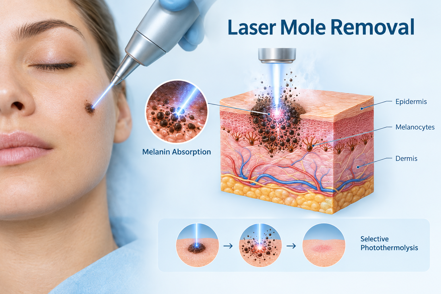

Laser mole removal is a minimally invasive outpatient procedure that uses focused light energy to break down pigmented cells within a mole. For the right candidate, it can produce a smooth cosmetic result with minimal downtime. However, safety depends heavily on whether the mole has been properly assessed beforehand, which laser technology is used, and whether the treating clinician has the appropriate expertise. This guide explains how the procedure works, who may be suitable, what the genuine risks are, and what questions to ask before booking a consultation.
Key Takeaways
- Laser mole removal is only appropriate for confirmed benign moles that have been assessed by a qualified clinician, ideally using dermoscopy.
- Because laser treatment destroys tissue rather than removing it intact, it does not allow for histopathological analysis, which means a suspicious mole must always be sent for biopsy via surgical excision instead.
- Common side effects include temporary redness, scabbing, and skin discoloration; post-inflammatory hyperpigmentation (PIH) is a particular concern for people with darker skin tones.
- Mole recurrence rates are higher with laser than with surgical excision, and incomplete removal is a documented risk for deeper moles.
- At-home laser devices are not a safe alternative and should be avoided entirely for mole treatment.
How Laser Mole Removal Works
Laser mole removal relies on a principle called selective photothermolysis, in which a laser emits a specific wavelength of light that is preferentially absorbed by melanin, the pigment responsible for a mole's colour. This absorption converts light energy into controlled heat, which destroys the melanocytes and pigmented tissue within the mole without affecting the surrounding skin to the same degree. The body then gradually eliminates the treated cells through its natural healing process.
The procedure is typically performed in an outpatient setting under local anesthesia or a topical numbing cream. Treatment time is usually short, often under thirty minutes for a small number of moles, and patients can generally return home the same day. Depending on the mole's size, depth, and pigmentation, more than one treatment session may be required to achieve a satisfactory result.
Types of Lasers Used for Mole Removal
Different laser technologies are suited to different mole characteristics and skin types. Understanding which device a clinic uses, and why, is a reasonable question to raise during a pre-treatment consultation.
- CO2 laser: An ablative laser that vaporises tissue layer by layer using photothermal energy. Effective for raised or thicker moles, but carries a higher risk of scarring and is generally better suited to lighter Fitzpatrick skin types (I–III).
- Erbium YAG laser: Also an ablative laser, but with a shorter wavelength that allows more precise tissue removal and less thermal spread to surrounding skin. Often preferred for superficial moles in areas requiring precision, such as the face.
- Q-switched Nd:YAG laser: A non-ablative laser that delivers nanosecond pulses targeting pigment without vaporising the surface tissue. Commonly used for flat, pigmented lesions and considered safer for medium skin tones.
- Picosecond (pico) laser: Delivers ultra-short pulses measured in picoseconds, producing a photoacoustic effect that shatters pigment with less thermal damage than older Q-switched devices. Often recommended for patients with darker skin tones where the risk of post-inflammatory hyperpigmentation is higher.
The choice of laser should be guided by the mole type, its depth, the patient's Fitzpatrick skin type, and the clinician's assessment. No single laser is universally optimal for every mole or every patient.
Is Laser Mole Removal Safe?
When performed by a board-certified dermatologist or appropriately qualified clinician using medically approved laser technology, laser mole removal is generally considered safe for confirmed benign lesions. The key word is confirmed. The procedure itself carries a well-understood risk profile, but the more significant safety concern is diagnostic: laser treatment destroys the mole's tissue, making it impossible to send a sample for histopathological analysis afterwards. If a mole that appeared benign turns out to contain early malignant cells, that opportunity for diagnosis is lost.
This is why a thorough pre-treatment assessment is not optional. A qualified clinician should evaluate every mole using dermoscopy, a non-invasive technique that uses magnification and polarised light to examine subsurface structures not visible to the naked eye. Dermoscopy allows the clinician to identify atypical features such as irregular borders, colour variation, or asymmetry that would indicate the mole should be excised and sent for biopsy rather than treated with a laser. Any mole with atypical features, any mole that has changed recently, or any mole in a patient with a personal or family history of melanoma should not be treated with laser removal.
The Diagnostic Safety Gap: Why This Matters
One of the most important safety considerations with laser mole removal is rarely explained clearly to patients. When a mole is surgically excised, the removed tissue is sent to a pathology laboratory for histopathological analysis. This process can detect early-stage malignant melanoma or other skin cancers even when the mole appeared clinically unremarkable. Laser treatment, by contrast, vaporises or fragments the tissue, leaving nothing intact to examine.
The practical consequence is this: if an early melanoma is present within a mole that is lasered without prior biopsy, the cancer may go undetected. The skin surface may appear to heal normally, while malignant cells that were not fully destroyed continue to develop beneath the surface. This is not a theoretical risk. It is the primary reason that clinical guidelines consistently recommend surgical excision for any mole that cannot be confidently confirmed as benign through dermoscopic assessment by an experienced clinician. Laser mole removal is a cosmetic procedure, not a diagnostic one, and it should only ever be used after the diagnostic question has been fully resolved.
Who May Be Suitable for Laser Mole Removal
Laser mole removal may be appropriate for patients whose moles meet specific clinical criteria. Suitable candidates generally include those with:
- Small, flat, or superficial moles confirmed as benign following clinical assessment
- Junctional nevi, which are moles where the pigmented cells sit at the junction between the epidermis and dermis, making them more accessible to laser energy
- Moles in cosmetically sensitive areas such as the face, nose, or ears where surgical excision might leave a more noticeable scar
- Lightly pigmented congenital moles that have been assessed and confirmed as non-cancerous
- Patients in good general health without active skin infections in the treatment area
Suitability must always be determined through a face-to-face consultation and dermoscopic assessment, not through photographs or self-assessment. A clinician who agrees to treat a mole without examining it in person is not following safe practice.
Who Should Not Have Laser Mole Removal
Laser mole removal is not appropriate for everyone, and certain situations make the procedure inadvisable or potentially dangerous:
- Moles with atypical features: Any mole showing asymmetry, irregular or poorly defined borders, multiple colours, or a diameter greater than six millimetres should be assessed for possible excision and biopsy, not laser treatment.
- Moles that have changed: A mole that has grown, changed colour, started bleeding, or become itchy requires urgent clinical evaluation, not cosmetic removal.
- Raised or deep dermal moles: Intradermal nevi extend deeper into the dermis and are less accessible to laser energy. Incomplete removal is more likely, and recurrence rates are higher.
- Patients with a history of melanoma: Any personal history of skin cancer requires specialist dermatological review before any mole removal procedure.
- Unassessed moles: No mole should be treated with a laser before it has been evaluated by a qualified clinician using appropriate diagnostic tools.
- Pregnancy: Elective cosmetic laser procedures are generally deferred during pregnancy.
Risks and Side Effects
Laser mole removal carries a range of potential side effects and risks that patients should understand before consenting to treatment. Most side effects are temporary and resolve during the healing process, but some complications can be longer-lasting.
Common and Expected Side Effects
- Redness and swelling: Normal immediately after treatment and usually resolves within a few days.
- Scabbing and crusting: Part of the natural wound healing process. Picking or scratching the area can increase scarring risk and infection.
- Temporary skin discoloration: The treated area may appear lighter or darker than the surrounding skin during healing.
Less Common but Significant Risks
- Post-inflammatory hyperpigmentation (PIH): Darkening of the treated skin, particularly common in patients with Fitzpatrick skin types IV–VI. PIH can persist for months and may require additional treatment.
- Hypopigmentation: Permanent lightening of the treated area, more likely with ablative lasers and in patients with naturally darker skin.
- Scarring: Atrophic (depressed) or hypertrophic (raised) scars can form, particularly if the mole was deep, if incorrect laser settings were used, or if aftercare instructions were not followed.
- Incomplete removal: If the mole's pigmented cells extend deeper than the laser can reach, residual cells may remain. This is a documented risk, particularly with raised or intradermal moles.
- Mole recurrence: Estimates suggest that mole recurrence following laser treatment may occur in approximately 10–20% of cases, a higher rate than with surgical excision. Recurrence does not automatically indicate malignancy, but any returning mole should be reassessed by a clinician.
- Infection: Any open wound carries a risk of infection. Signs include spreading redness, warmth, pus, or fever above 38°C (100.4°F), all of which require prompt medical attention.
- Delayed melanoma diagnosis: As discussed above, the inability to perform histopathological analysis on lasered tissue is a serious safety consideration if the pre-treatment assessment was inadequate.
Skin Type and Laser Safety
A patient's Fitzpatrick skin type is a clinically important factor in determining which laser is safest and what risks are most relevant. The Fitzpatrick scale classifies skin from type I (very fair, always burns) through to type VI (deeply pigmented, never burns). Ablative lasers such as the CO2 laser are generally better tolerated by patients with Fitzpatrick types I–III, where the contrast between the mole's melanin and the surrounding skin is greater and the risk of collateral pigment disruption is lower.
Patients with Fitzpatrick types IV–VI have a significantly higher risk of post-inflammatory hyperpigmentation following ablative laser treatment. For these patients, a picosecond laser or Q-switched Nd:YAG laser may be a safer option, as these devices produce less thermal damage to surrounding tissue. Even with these technologies, the risk of PIH is not eliminated, and realistic expectations should be discussed during the pre-treatment consultation. Clinics that do not ask about skin type or that offer a single laser for all patients regardless of skin tone may not be following best practice.
Laser Mole Removal vs. Surgical Excision
Surgical excision remains the clinical gold standard for mole removal, particularly for any mole that requires pathological examination. Understanding the differences between the two approaches helps patients make an informed decision in consultation with their clinician.
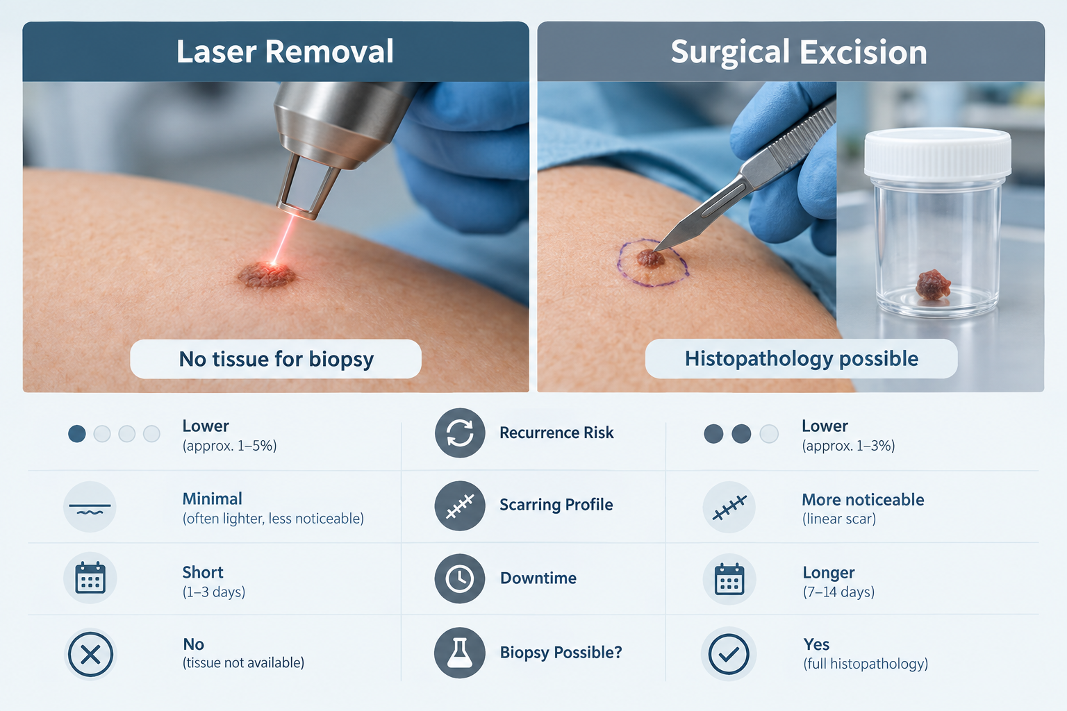
| Factor | Laser Mole Removal | Surgical Excision |
|---|---|---|
| Tissue for biopsy | No — tissue is destroyed | Yes — specimen sent to pathology |
| Best suited for | Small, flat, confirmed benign moles | Any mole, especially suspicious or raised ones |
| Scarring profile | Minimal scarring possible; risk of PIH or hypopigmentation | Linear scar likely; fades over time |
| Recurrence rate | Higher (estimated 10–20%) | Lower when margins are clear |
| Downtime | Typically 1–3 weeks healing | Typically 1–4 weeks depending on size |
| Sessions required | May require multiple sessions | Usually one procedure |
| Suitable for deep moles | Limited — incomplete removal risk | Yes — full depth removal possible |
| Cosmetic outcome on face | Can be excellent for superficial moles | Linear scar; may be less cosmetically ideal in some areas |
The choice between laser and surgical excision should be guided by the mole's clinical characteristics, not by patient preference alone. A clinician who recommends laser removal without first ruling out atypical features is not following safe practice.
What to Expect During and After Treatment
During the Procedure
Laser mole removal is performed in a clinical setting. The skin is cleaned and a local anesthetic or numbing cream is applied to minimize discomfort. Protective eyewear is worn by both the patient and the clinician. The laser is then applied to the mole in controlled pulses. Most patients describe the sensation as a mild snapping or warmth. The procedure itself is usually brief, though the exact duration depends on the number and size of moles being treated.
Recovery Timeline
- Days 0–3: Redness, mild swelling, and tenderness are normal. A scab or crust will begin to form over the treated area.
- Days 3–21: The scab gradually lifts as the skin heals beneath it. The area may appear pink or slightly discolored. Itching is common but scratching should be avoided.
- Three weeks to several months: Skin tone and texture continue to normalize. Any residual redness or discoloration typically fades during this period, though PIH may take longer to resolve in some patients.
Aftercare Essentials
- Keep the treated area clean using mild soap or a fragrance-free cleanser.
- Apply an antibiotic or healing ointment as directed by your clinician.
- Do not pick, scratch, or rub the scab — allow it to fall away naturally.
- Avoid direct sun exposure on the treated area during healing.
- Apply a broad-spectrum SPF 30 or higher sunscreen once the skin has healed, and continue daily sun protection for several months to reduce the risk of PIH.
- Attend all scheduled follow-up appointments so your clinician can monitor healing and identify any complications early.
- Seek medical advice promptly if you notice spreading redness, pus, fever, or any signs of infection.
The Danger of At-Home Laser Devices
Consumer-grade at-home laser devices marketed for skin concerns are not appropriate for mole removal and should not be used for this purpose. The Skin Cancer Foundation has warned that attempting to treat moles with at-home devices can alter the appearance of mole cells, potentially masking early signs of malignancy and delaying a diagnosis of melanoma. Unlike medically approved laser technology used in a clinical setting, at-home devices are not calibrated for mole tissue, are not preceded by dermoscopic assessment, and are operated without clinical oversight.
DIY mole removal using any method — including at-home laser devices, over-the-counter creams, or physical removal — carries serious risks and should be avoided entirely. If a mole is causing concern, the appropriate first step is always a consultation with a qualified clinician.
Questions to Ask Your Provider Before Booking
Choosing a qualified and experienced provider is one of the most important safety decisions a patient can make. Before committing to treatment, consider asking the following:
- Will you assess my mole using dermoscopy before recommending laser treatment?
- How do you determine whether a mole is suitable for laser removal or should be excised instead?
- Which laser technology will you use, and why is it appropriate for my mole and skin type?
- What is your experience with treating patients of my Fitzpatrick skin type?
- What is the realistic risk of mole recurrence, and what happens if the mole returns?
- What aftercare support do you provide, and how do I contact you if I have concerns during healing?
- Are you a board-certified dermatologist or do you work under the supervision of one?
A clinician who is unwilling or unable to answer these questions clearly is not the right choice for this procedure.
Frequently Asked Questions
Can all moles be removed by laser?
No. Laser mole removal is only appropriate for small, flat, superficial moles that have been confirmed as benign through clinical assessment. Raised moles, deep dermal moles, and any mole with atypical features are generally not suitable candidates. Suspicious moles must be surgically excised so the tissue can be examined pathologically.
Does laser mole removal hurt?
Most patients experience minimal discomfort during the procedure because local anesthesia or a numbing cream is applied beforehand. Some describe a mild snapping sensation or warmth during laser pulses. Mild soreness and tenderness in the treated area are normal for a few days afterwards.
Will laser mole removal leave a scar?
Scarring is possible but not inevitable. The risk depends on the mole's depth, the laser used, the clinician's technique, and how well aftercare instructions are followed. Superficial moles treated with appropriate laser settings by an experienced clinician often heal with minimal visible scarring, though some degree of skin texture or colour change is possible.
Can a mole grow back after laser treatment?
Yes. Mole recurrence is a documented limitation of laser removal, with estimates suggesting it may occur in roughly 10–20% of cases. Recurrence is more likely with deeper moles where incomplete removal is possible. If a treated mole returns, it should be reassessed by a clinician rather than retreated without evaluation, as any returning lesion warrants fresh dermoscopic assessment.
Is laser mole removal covered by insurance?
In most cases, laser mole removal performed for cosmetic reasons is not covered by health insurance. The procedure is typically classified as an elective cosmetic treatment. Costs vary depending on the clinic, the number of moles treated, and the laser technology used. If a mole requires removal for medical reasons, coverage may differ, and it is worth checking with your insurer and clinician.
How many sessions will I need?
This depends on the mole's size, depth, and pigmentation. Some superficial moles respond fully in a single session, while others may require two or more treatment sessions to achieve a satisfactory result. Your clinician should give you a realistic estimate during the pre-treatment consultation.
Making a Safe and Informed Decision About Laser Mole Removal
Laser mole removal can be a safe and effective option for the right patient with the right mole, assessed by the right clinician. The procedure works well for small, flat, confirmed benign lesions in patients who have undergone a proper dermoscopic evaluation and who understand the realistic outcomes, including the possibility of requiring multiple sessions, the risk of PIH in darker skin tones, and the higher recurrence rate compared to surgical excision.
The procedure is not appropriate as a shortcut to avoid a proper clinical assessment, and it is never appropriate for moles that have not been evaluated for atypical features. The inability to perform histopathological analysis on lasered tissue is a genuine and serious limitation that should be part of every informed consent discussion. Patients with darker skin tones should specifically discuss PIH risk and laser selection with their clinician before proceeding.
If you are considering laser mole removal, the most important first step is a consultation with a board-certified dermatologist or qualified clinician who uses dermoscopy as part of their assessment process. A thorough pre-treatment evaluation protects your health and gives you the clearest picture of whether laser removal is genuinely the right approach for your specific mole.
