
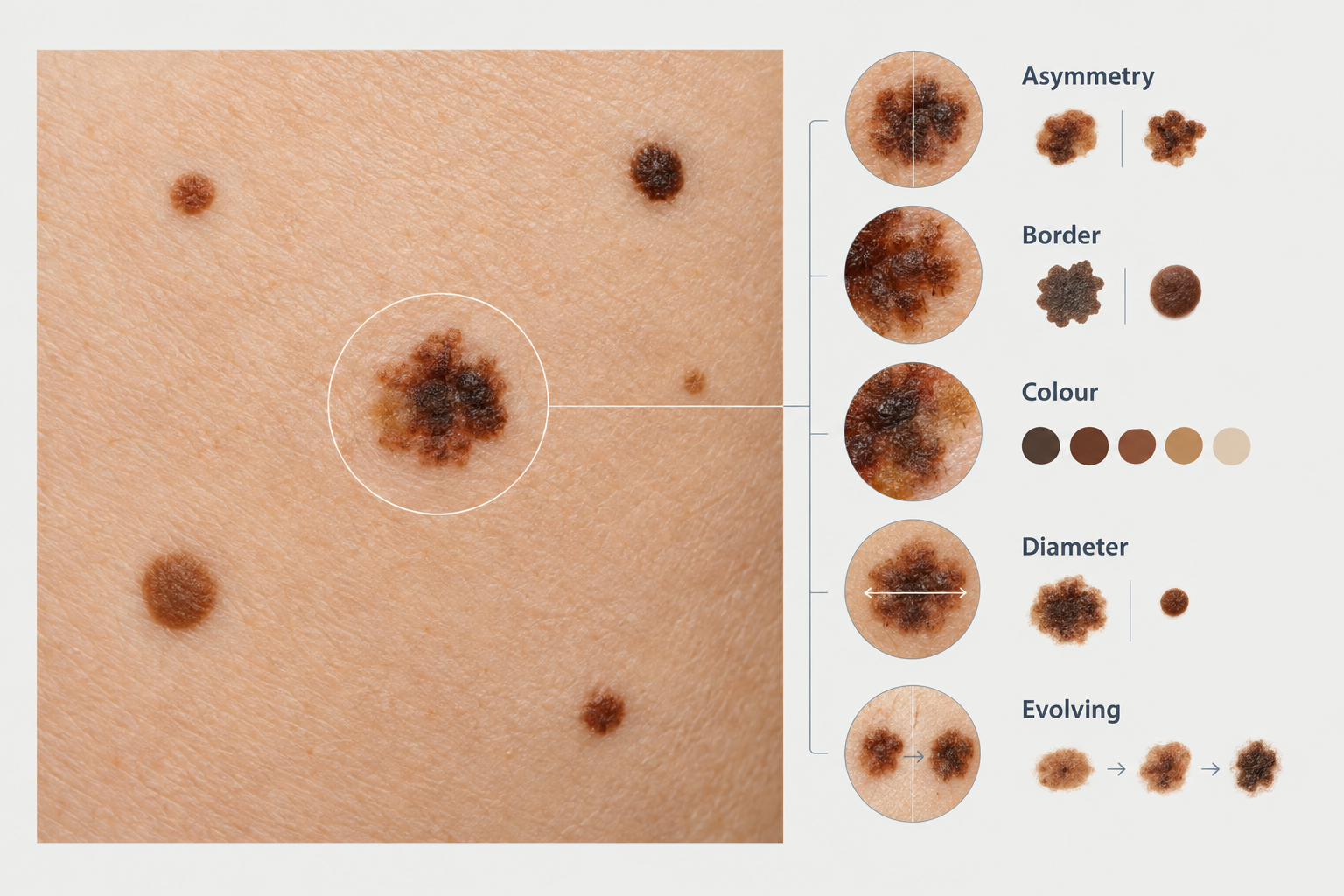

Most people have moles, and the vast majority are completely harmless. Knowing when to get moles checked, however, is one of the most practical steps you can take for your long-term skin health. Changes in size, shape, colour, or texture can occasionally signal something that warrants professional assessment, and catching any concern early generally leads to better outcomes. This article explains the warning signs to watch for, how often routine checks are recommended, what a professional mole assessment involves, and when you should seek an appointment without delay.
Key Takeaways
- Most moles are benign, but any mole that changes in size, shape, colour, or texture should be assessed by a qualified clinician promptly.
- The ABCDE criteria — Asymmetry, Border, Colour, Diameter, and Evolving — provide a practical self-check framework, but they do not replace professional examination.
- Annual skin checks are commonly recommended for people with a higher number of moles, a personal or family history of skin cancer, or significant past sun exposure.
- Dermoscopy, a non-invasive magnification technique, allows clinicians to examine mole structure in detail that is not visible to the naked eye.
- If a mole bleeds, itches persistently, becomes raised, or develops an irregular border, seek a professional assessment rather than waiting for a routine appointment.
What Is a Mole?
Moles, known medically as melanocytic naevi, are clusters of pigment-producing cells called melanocytes that grow in the skin. They can appear anywhere on the body, including the scalp, between the toes, and under the nails. Most moles develop during childhood and adolescence, though new moles can appear in adulthood. The average adult has between 10 and 40 moles, and having a higher number — generally considered to be more than 50 — is associated with a modestly increased risk of melanoma, the most serious form of skin cancer.
Moles vary considerably in appearance. They may be flat or raised, smooth or rough, and range in colour from pale pink to dark brown or black. Some have hair growing from them. These variations are normal. What matters clinically is not how a mole looks at a single point in time, but whether it is changing, and whether any features suggest it may need closer investigation.
The ABCDE Warning Signs
The ABCDE criteria were developed to help both patients and clinicians identify moles that may warrant further evaluation. Understanding these criteria can help you decide when to get a mole checked between routine appointments.
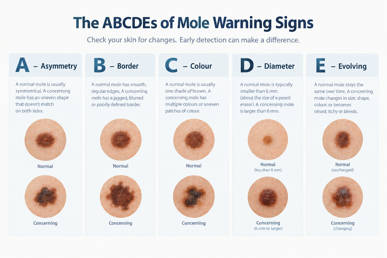
- Asymmetry: A typical benign mole is roughly symmetrical — if you drew a line through the centre, both halves would look similar. A mole where one half looks noticeably different from the other may be worth assessing.
- Border: Benign moles tend to have smooth, well-defined edges. Irregular, ragged, notched, or blurred borders can be a reason to seek professional review.
- Colour: A uniform colour — whether brown, tan, or black — is generally reassuring. Moles that contain multiple shades, or that include areas of red, white, or blue-black, may need examination.
- Diameter: Moles larger than approximately 6 millimetres — roughly the size of a pencil eraser — are traditionally flagged for review, though melanomas can be smaller, and many large moles are benign.
- Evolving: This is arguably the most important criterion. Any mole that is changing — in size, shape, colour, or texture — or that develops new symptoms such as itching, bleeding, or crusting, should be assessed by a clinician.
The ABCDE framework is a useful starting point for self-examination, but it has limitations. Some melanomas do not fit neatly into these categories, and some benign moles may appear to tick several boxes. Professional assessment with dermoscopy provides considerably more diagnostic information than visual inspection alone.
When to Get a Mole Checked Without Delay
Certain changes should prompt you to seek a professional assessment promptly rather than waiting for a scheduled skin check. These include a mole that bleeds without injury, itches persistently, becomes inflamed or develops a surrounding redness, crusts or scabs repeatedly, or changes noticeably over a period of weeks rather than years. A new dark lesion that appears in adulthood — particularly after the age of 40 — also warrants attention, as most new moles in adults are benign but some may not be.
Pain in or around a mole is another reason to seek assessment. While most moles are painless, a mole that becomes tender or sore without an obvious cause such as friction or trauma should be reviewed. Similarly, if a previously flat mole becomes raised or develops a lump within it, this change in texture is worth discussing with a clinician.
It is worth emphasising that experiencing one or more of these changes does not mean a mole is cancerous. Many benign conditions, including seborrhoeic keratoses, dermatofibromas, and irritated moles, can produce similar symptoms. The purpose of seeking assessment is to obtain an accurate diagnosis, not to assume the worst.
How Often Should Moles Be Checked Professionally?
For most people with a small number of typical-looking moles and no significant risk factors, a professional skin check every one to two years is a reasonable approach, though recommendations vary between healthcare systems and individual clinical circumstances. Your clinician may suggest a different interval based on your personal risk profile.
More frequent monitoring — often annually or even every six months — is generally recommended for people who have a larger number of moles, a personal history of melanoma or other skin cancers, a first-degree relative who has had melanoma, a history of significant or repeated sunburn, a history of using sunbeds, fair skin that burns easily, or a condition that affects immune function. People with atypical mole syndrome, sometimes called dysplastic naevus syndrome, in which moles are numerous and irregular in appearance, are typically placed on a structured surveillance programme.
Between professional checks, monthly self-examination is widely encouraged. Becoming familiar with your own moles makes it easier to notice when something has changed. Photographs taken in consistent lighting can be a practical way to track moles over time, though they are not a substitute for clinical assessment with dermoscopy.
Who Is at Higher Risk and May Need More Frequent Checks?
Skin cancer risk is influenced by a combination of genetic, environmental, and skin-type factors. People with fair skin, light eyes, and red or blonde hair tend to have less melanin — the pigment that provides some protection against ultraviolet radiation — and may burn more easily. Cumulative sun exposure over a lifetime, particularly with repeated episodes of sunburn, is one of the most significant modifiable risk factors for melanoma and other skin cancers.
A family history of melanoma increases individual risk, particularly if a first-degree relative was affected. Certain inherited gene variants, including mutations in the CDKN2A gene, are associated with a substantially higher lifetime risk of melanoma, and genetic counselling may be appropriate in some families with multiple affected members.
People who are immunosuppressed — for example, following organ transplantation or due to certain medical conditions or medications — have a higher risk of skin cancers, including squamous cell carcinoma, and may benefit from more frequent dermatological review. If you are unsure whether your personal circumstances place you in a higher-risk category, discussing this with your GP or a dermatologist is a sensible starting point.
What Happens During a Professional Mole Assessment?
A professional mole check typically begins with a clinical history, during which the clinician will ask about any moles you are concerned about, how long you have had them, whether they have changed, and relevant background information such as your skin type, sun exposure history, and any personal or family history of skin cancer. This context helps the clinician prioritise which lesions to examine most carefully.
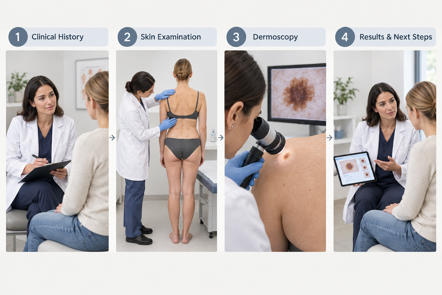
The physical examination involves inspecting the skin systematically, often from head to toe, to assess all visible moles and other pigmented lesions. Clinicians trained in skin cancer assessment use dermoscopy — a handheld device that illuminates and magnifies the skin surface — to examine the internal structure of moles in detail. Dermoscopy allows assessment of features such as pigment network patterns, vascular structures, and architectural symmetry that are not visible to the naked eye, and it significantly improves diagnostic accuracy compared with naked-eye examination alone.
Some clinics use total body photography or digital dermoscopy systems that store images of moles over time, enabling precise comparison at future appointments. This approach is particularly useful for monitoring patients with a large number of moles or atypical lesions.
If a mole raises concern during assessment, the clinician may recommend a biopsy — the removal of a small tissue sample for laboratory analysis — or referral to a dermatologist or plastic surgeon for excision. A recommendation to remove a mole does not automatically mean it is malignant; excision may be advised to obtain a definitive diagnosis or because the mole has features that make monitoring alone insufficient.
Types of Skin Lesions That Are Sometimes Confused with Moles
Not every pigmented spot on the skin is a mole. Several common benign conditions can resemble moles and are sometimes a source of unnecessary concern, while other lesions that do not look like typical moles can occasionally be more significant. Understanding the range of lesions that may be encountered during a skin check can help set realistic expectations.
Seborrhoeic keratoses are very common benign growths that often appear in middle age and beyond. They can be brown, black, or pale, and may have a rough, warty, or stuck-on appearance. They are not related to sun damage and carry no malignant potential, though they can sometimes be difficult to distinguish from melanoma without dermoscopy. Dermatofibromas are firm, slightly raised nodules that are usually brown and often appear on the legs. Solar lentigines — commonly called age spots or liver spots — are flat, pigmented areas caused by cumulative sun exposure and are benign, though they can occasionally be confused with lentigo maligna, an early form of melanoma that develops slowly on sun-damaged skin.
Basal cell carcinoma and squamous cell carcinoma, the two most common forms of skin cancer, do not always arise from existing moles and may not be pigmented. A pearly or translucent bump, a persistent scaly patch, or a sore that does not heal are all reasons to seek professional assessment, even if the lesion does not look like a typical mole.
Self-Examination: A Practical Approach
Regular self-examination of the skin is a practical complement to professional checks. Performing a systematic self-check once a month, in good lighting and using a full-length mirror and a hand mirror for harder-to-see areas, allows you to become familiar with your own skin and notice changes more readily. A partner or family member can help examine areas such as the scalp and back.
When examining a mole, consider whether it looks the same as it did last month. If you are unsure, a photograph taken in consistent lighting and from the same angle can serve as a reference point. Note any moles that feel different — itchy, tender, or rough — as well as those that look different. If you notice a change that concerns you, seek professional assessment rather than monitoring it yourself for several more months.
Self-examination is not a substitute for professional assessment and has recognised limitations. Studies suggest that patients often miss lesions in difficult-to-see locations and may not reliably identify early melanomas. Professional dermoscopy remains the standard for accurate mole evaluation.
Risks of Delaying a Mole Check
Melanoma is one of the most serious skin cancers because of its potential to spread to other parts of the body if not detected and treated early. When melanoma is identified at an early stage — while it is still confined to the outer layers of the skin — the prognosis is generally favourable, and treatment is typically straightforward surgical excision. As melanoma grows deeper into the skin or spreads to lymph nodes or distant organs, treatment becomes more complex and outcomes less predictable.
Delaying assessment of a changing mole does not guarantee a worse outcome, and the majority of moles that change are not melanoma. However, the potential consequences of missing an early melanoma mean that erring on the side of seeking professional review is generally the more prudent approach. There is no clinical downside to having a mole assessed and being reassured that it is benign.
Frequently Asked Questions
Can a new mole appear in adulthood?
New moles can appear in adulthood, and most are benign. However, a new pigmented lesion appearing after the age of 40, or any new lesion that grows quickly or looks unusual, is worth having assessed by a clinician rather than assumed to be harmless.
Is it normal for moles to change during pregnancy?
Hormonal changes during pregnancy can cause some moles to darken or become slightly larger. These changes are usually benign, but any mole that changes significantly during pregnancy should still be assessed by a clinician, as pregnancy does not protect against melanoma.
Do moles need to be removed if they are benign?
Benign moles do not need to be removed for medical reasons, though some people choose to have moles removed for cosmetic reasons or because they are in a location that causes repeated irritation. If removal is desired, this should be performed by a qualified clinician and the removed tissue should be sent for histological analysis.
Can sunscreen reduce the risk of mole changes?
Regular use of broad-spectrum sunscreen with a high SPF, combined with protective clothing and avoiding peak sun hours, is associated with a reduced risk of new mole development and may help reduce the risk of sun-related skin damage more broadly. Sunscreen does not eliminate risk entirely, and regular skin checks remain important regardless of sun protection habits.
What is the difference between a mole check at a GP and a dermatologist?
A GP can perform an initial assessment and refer you for further evaluation if needed. A dermatologist has specialist training in skin conditions and is more likely to use dermoscopy routinely. In some settings, specialist skin cancer clinics or aesthetic medicine practitioners with specific training in dermoscopy offer dedicated mole assessment services. The most important factor is that the clinician performing the assessment has appropriate training and access to dermoscopy.
How long does a mole check appointment take?
A focused assessment of one or two specific moles may take 10 to 15 minutes. A full-body skin check, in which all visible moles and lesions are systematically examined, typically takes 20 to 45 minutes depending on the number of lesions and the thoroughness of the assessment.
What happens if a mole needs to be removed?
Mole removal is usually performed under local anaesthetic as a minor surgical procedure. The mole and a small margin of surrounding skin are excised, and the wound is closed with sutures. The removed tissue is sent to a laboratory for histological analysis to confirm the diagnosis. Results are typically available within one to two weeks, and the clinician will discuss the findings and any further steps required.
When to Act on a Mole Concern
The practical decision about when to get moles checked comes down to a straightforward principle: if something has changed, or if you are uncertain, seek professional assessment. Waiting to see whether a changing mole resolves on its own is rarely the right approach, because benign changes do not require treatment and concerning changes benefit from early attention. There is no clinical cost to seeking reassurance.
For people without specific risk factors, incorporating a professional skin check into routine healthcare every one to two years is a reasonable habit. For those with a higher number of moles, a relevant personal or family history, or significant past sun exposure, more frequent structured monitoring with dermoscopy provides a meaningful safety net. Between appointments, monthly self-examination and sensible sun protection habits remain practical and worthwhile steps. If you have a mole that concerns you, the most useful thing you can do is book an assessment with a qualified clinician rather than searching for reassurance online.
