
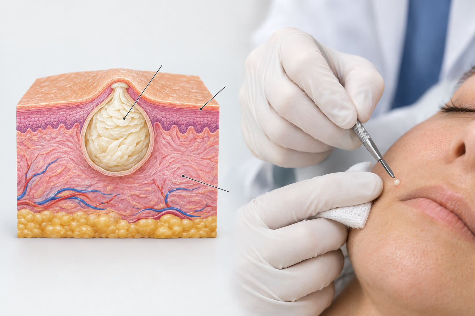

Milia are small, keratin-filled cysts that sit just beneath the skin's surface, appearing as firm white or yellowish bumps most commonly around the eyes, nose, and cheeks. While they are entirely harmless, many people seek professional milia removal because the bumps are persistent and do not resolve with standard skincare alone. This article explains what milia are, why they form, which removal methods are available, what each procedure involves, and how to choose a safe approach with the help of a qualified practitioner.
Key Takeaways
- Milia are benign keratin cysts that rarely disappear on their own and typically require professional treatment for reliable removal.
- Several safe removal methods exist, including manual extraction, electrocautery, laser ablation, and topical retinoids, each with different suitability profiles and recovery expectations.
- Attempting to squeeze or pierce milia at home carries a real risk of scarring, infection, and post-inflammatory hyperpigmentation.
- A clinical assessment is important before treatment, as some skin lesions that resemble milia may require a different diagnosis and management approach.
- Results after professional removal are generally good, but milia can recur, particularly if underlying causes such as heavy occlusive skincare or sun damage are not addressed.
What Are Milia and Why Do They Form?
Milia (singular: milium) are tiny epidermal inclusion cysts, typically one to two millimetres in diameter, filled with keratin — the same structural protein found in hair and nails. Unlike whiteheads, milia have no pore opening, which is why they cannot be cleared by washing or gentle exfoliation. The overlying skin is intact, trapping the keratin beneath the surface.
Milia are classified into two main categories. Primary milia arise spontaneously, often in newborns (where they resolve without treatment within weeks) or in adults around the eyelids and cheeks. Secondary milia develop as a consequence of skin trauma, blistering conditions, prolonged use of heavy occlusive creams, topical corticosteroid use, or sun damage that disrupts normal skin cell turnover. Understanding which type is present helps guide the most appropriate treatment approach.
Common contributing factors in adults include the use of thick emollient-rich moisturisers or eye creams that block follicular openings, cumulative ultraviolet damage that thickens the outer skin layer, and certain skin conditions such as bullous pemphigoid or porphyria cutanea tarda. In many cases, however, no single clear cause is identified.
Can Milia Resolve Without Treatment?
Primary milia in newborns typically clear within four to six weeks without any intervention. In adults, primary milia around the eyes and cheeks are far less likely to resolve spontaneously and may persist for months or years. Secondary milia, which form after skin injury or prolonged use of occlusive products, may gradually improve once the triggering factor is removed, but this is not guaranteed and can take considerable time.
Switching to lighter, non-comedogenic skincare formulations and incorporating a gentle retinoid or exfoliating acid into a routine may support natural skin cell turnover and reduce the likelihood of new milia forming. However, these measures are generally more useful for prevention than for clearing established milia, which usually require a physical or procedural intervention to remove reliably.
Professional Milia Removal Methods
Several clinically recognised techniques are used for safe milia removal. The most appropriate method depends on the number of milia present, their location, the patient's skin type, and the practitioner's assessment. Each approach has a distinct mechanism, recovery profile, and risk consideration.
Manual Extraction
Manual extraction is the most widely used method for milia removal and is considered the standard first-line approach in most clinical settings. The practitioner uses a sterile lancet or fine needle to create a small opening in the overlying skin, then gently expresses the keratin contents using a comedone extractor or light manual pressure. The procedure is typically quick, taking only a few minutes for a small number of milia, and requires no anaesthetic for most patients, though a topical numbing cream may be applied in sensitive areas such as the lower eyelid.
Manual extraction is well-suited to isolated or small clusters of milia and carries a low risk of scarring when performed by a trained practitioner using sterile technique. The main risks include temporary redness, minor bruising, and a small risk of post-inflammatory hyperpigmentation, particularly in individuals with darker skin tones. Attempting manual extraction at home without sterile equipment significantly increases the risk of infection and scarring, which is why professional treatment is strongly recommended.
Electrocautery and Diathermy
Electrocautery uses a fine electrical current to precisely ablate the milium and its contents. A small probe delivers controlled heat to the cyst, effectively destroying the keratin plug and the cyst wall. This method is particularly useful for milia that are deeply seated or located in areas where manual extraction is technically difficult. Electrocautery is generally well-tolerated, though a brief stinging sensation is common during the procedure. A small crust typically forms at the treatment site and resolves within one to two weeks.
The risk of post-treatment hyperpigmentation or hypopigmentation is slightly higher with electrocautery compared to manual extraction, particularly in patients with Fitzpatrick skin types IV to VI. A patch test or test treatment on a small area may be advisable before treating multiple milia in patients with darker skin tones.
Laser Ablation
Laser treatment for milia typically uses a carbon dioxide (CO2) laser or an erbium:YAG laser to precisely vaporise the cyst contents and the surrounding tissue. Laser ablation offers a high degree of precision and is particularly useful for milia located very close to the eyelid margin, where manual extraction carries a higher risk of injury to the delicate periorbital skin. The procedure is usually performed under topical anaesthesia, and protective eyewear is essential when treating the periorbital area.
Recovery after laser milia removal involves a short period of redness and crusting at the treated sites, typically resolving within one to two weeks. The risk of temporary pigment changes is present, and patients with a tendency toward post-inflammatory hyperpigmentation may require a longer recovery period or pre-treatment skin preparation. Laser treatment is generally reserved for cases where other methods are less suitable or where a larger number of milia need to be addressed efficiently.
Chemical Peels and Topical Retinoids
Chemical peels using alpha-hydroxy acids (AHAs) such as glycolic acid, or beta-hydroxy acids such as salicylic acid, can support the gradual breakdown of the keratin plugs responsible for milia by accelerating epidermal cell turnover. Superficial peels are not typically sufficient to remove established milia on their own but may be used as an adjunct to other treatments or as a preventive measure to reduce recurrence. Medium-depth peels may have a more meaningful effect on milia but carry a higher risk of downtime and pigment changes.
Topical retinoids, including tretinoin and adapalene, work by normalising keratinocyte differentiation and promoting regular skin cell shedding, which can help prevent new milia from forming and may gradually reduce existing ones over several months of consistent use. Retinoids are available in both prescription and over-the-counter formulations, and their use should be discussed with a qualified practitioner, particularly for application near the eyes where skin is thin and sensitive.
Cryotherapy
Cryotherapy involves the application of liquid nitrogen to freeze and destroy the milium. While cryotherapy is commonly used for other benign skin lesions such as viral warts and seborrhoeic keratoses, its use for milia is less common because the precision required to treat small, superficial cysts without damaging surrounding skin can be challenging. There is also a meaningful risk of post-treatment hypopigmentation, particularly in patients with darker skin tones. Cryotherapy for milia is generally considered when other methods are not suitable or available.
What Happens During a Milia Removal Appointment?
A professional milia removal appointment typically begins with a clinical assessment of the skin lesions to confirm the diagnosis. Some conditions, including closed comedones, syringomas, sebaceous hyperplasia, and xanthelasma, can resemble milia and require a different management approach. A trained practitioner will examine the lesions carefully and may ask about skincare habits, medical history, and any previous skin treatments before proceeding.
Once milia removal is confirmed as appropriate, the skin is cleansed and, if necessary, a topical anaesthetic cream is applied and left for a short period. The chosen removal technique is then performed, with the practitioner working methodically through each milium. Most appointments for a small number of milia take between fifteen and thirty minutes. Immediately after treatment, the skin may appear slightly red or swollen at the treated sites, and small crusts may form depending on the method used.
Recovery and Aftercare
Recovery after professional milia removal is generally straightforward. Following manual extraction, most patients experience mild redness that settles within a few hours to a day. After electrocautery or laser treatment, small crusts form at each treated site and should be left to heal naturally without picking, as premature removal of crusts increases the risk of scarring and pigment changes.
Aftercare guidance typically includes keeping the treated area clean, avoiding heavy occlusive products on the treated skin while it heals, applying a broad-spectrum SPF 30 or higher sunscreen daily to protect healing skin from ultraviolet exposure, and avoiding active exfoliants such as AHAs, BHAs, and retinoids on the treated area until the skin has fully recovered. Sun protection is particularly important in the weeks following treatment to minimise the risk of post-inflammatory hyperpigmentation.
Most patients can return to normal daily activities immediately or within one to two days, depending on the extent of treatment and the method used. Makeup can usually be applied once any crusting has resolved, which is typically within five to ten days for electrocautery or laser treatments.
Risks and Side Effects of Milia Removal
Professional milia removal is generally considered low-risk when performed by a trained and experienced practitioner using appropriate technique and sterile equipment. However, as with any skin procedure, potential side effects and complications should be understood before treatment.
- Temporary redness and swelling: Common after all methods and usually resolves within hours to a few days.
- Crusting: Expected after electrocautery and laser treatment; resolves within one to two weeks.
- Post-inflammatory hyperpigmentation (PIH): Darkening of the skin at treated sites, more common in individuals with Fitzpatrick skin types III to VI. Usually temporary but may take several weeks or months to fade.
- Hypopigmentation: Lightening of the skin at treated sites, more commonly associated with cryotherapy and laser treatments. May be prolonged in some cases.
- Scarring: Uncommon with professional treatment but possible, particularly if aftercare instructions are not followed or if the skin is manipulated during healing.
- Infection: Rare when sterile technique is used; risk increases significantly with home attempts at removal.
- Incomplete removal or recurrence: Some milia may not be fully cleared in a single session, and new milia can develop over time, particularly if contributing factors are not addressed.
Who May Be Suitable for Professional Milia Removal?
Most adults with confirmed milia who are bothered by their appearance are potential candidates for professional removal. Suitability for a specific method depends on several factors that a practitioner will assess during consultation.
Individuals with a history of keloid scarring may be at higher risk of adverse outcomes with more invasive methods such as electrocautery or laser and may be better suited to manual extraction or topical approaches. Patients with active skin infections, open wounds, or inflammatory skin conditions in the treatment area should wait until these have resolved before undergoing removal. Those taking oral isotretinoin or who have recently completed a course should inform their practitioner, as wound healing may be affected.
Pregnancy is a relevant consideration for some treatment methods, particularly those involving chemical agents or certain laser wavelengths. A thorough medical history and discussion of current medications, including topical treatments, is an important part of the pre-treatment assessment. Patients with very dark skin tones should discuss the risk of pigment changes with their practitioner before choosing a method, as some techniques carry a higher risk of PIH or hypopigmentation in this group.
Preventing Milia Recurrence
Milia can recur after successful removal, particularly if the factors that contributed to their formation are not modified. Several practical measures may help reduce the likelihood of new milia developing.
- Switching to lighter, non-comedogenic moisturisers and eye creams, particularly around the periorbital area where milia are most common.
- Incorporating a low-concentration retinoid or AHA-based product into a regular skincare routine to support normal skin cell turnover, after discussing suitability with a practitioner.
- Using a broad-spectrum sunscreen daily to limit cumulative ultraviolet damage, which can thicken the outer skin layer and impair normal desquamation.
- Avoiding prolonged use of potent topical corticosteroids on the face unless medically necessary and supervised.
- Attending periodic skin reviews with a practitioner if milia recur frequently, to assess whether an underlying skin condition or skincare habit is contributing.
Alternatives to Professional Removal
For patients who prefer a non-procedural approach or who have very mild milia, some alternatives may be worth discussing with a practitioner, though their effectiveness for established milia is generally more limited than professional removal techniques.
Consistent use of a topical retinoid such as tretinoin or adapalene over several months may gradually reduce existing milia and help prevent new ones from forming by normalising keratinocyte turnover. Regular use of a gentle AHA-based exfoliant, such as a glycolic acid toner or serum, may support the same process. These approaches require patience and consistent use, and results are not guaranteed for all patients.
Professional facial treatments such as microdermabrasion or superficial chemical peels may complement other approaches by improving overall skin texture and supporting cell renewal, but are unlikely to clear established milia on their own. These options may be more useful as maintenance treatments after professional milia removal to reduce recurrence.
Frequently Asked Questions
Is milia removal painful?
Manual extraction of milia typically causes minimal discomfort, described by most patients as a brief sharp sensation. Electrocautery and laser treatments may cause a stinging or heat sensation, which is usually well-tolerated. Topical anaesthetic cream can be applied before treatment to improve comfort, particularly for milia near the eyelids.
How many sessions are needed to remove milia?
Many milia can be removed in a single appointment. However, if a large number of milia are present, or if new milia develop over time, additional sessions may be needed. A practitioner can advise on a realistic treatment plan based on the number and distribution of milia present.
Can I remove milia at home safely?
Attempting to pierce or squeeze milia at home is not recommended. Without sterile equipment and proper technique, home attempts carry a meaningful risk of infection, scarring, and post-inflammatory hyperpigmentation. Professional treatment by a trained practitioner is the safest and most effective approach.
Will milia come back after removal?
Milia can recur after professional removal, particularly if contributing factors such as heavy occlusive skincare, sun damage, or certain skin conditions are not addressed. Adopting appropriate skincare habits and attending follow-up reviews can help reduce the likelihood of recurrence.
Are milia the same as whiteheads?
No. Milia and whiteheads are distinct conditions. Whiteheads are a form of acne comedone with a pore opening that can be cleared with exfoliation or topical treatments. Milia are keratin-filled cysts with no pore opening, which is why they do not respond to standard acne treatments and require a different removal approach.
Is milia removal suitable for the area around the eyes?
Yes, milia around the eyes are among the most common locations treated. The periorbital area requires particular care due to the thinness and sensitivity of the skin. Manual extraction and laser ablation are both used in this area, with appropriate protective measures such as eye shields when laser treatment is performed near the eyelid margin.
How long does it take to see results after milia removal?
Results from manual extraction are typically visible immediately, as the milia are physically removed during the appointment. After electrocautery or laser treatment, small crusts form at the treated sites and the final result becomes apparent once these have healed, usually within one to two weeks.
Deciding on the Right Milia Removal Approach
Milia removal is a straightforward and generally low-risk procedure when carried out by a qualified practitioner, but the most appropriate method varies depending on the number of milia, their location, the patient's skin type, and individual medical history. Manual extraction remains the most widely used and accessible option for most patients, while electrocautery and laser ablation offer useful alternatives for deeper or more numerous milia, or for cases where precision is particularly important.
Before proceeding with any removal method, a clinical assessment is important to confirm the diagnosis and rule out other skin lesions that may look similar but require different management. Addressing contributing factors — such as switching to lighter skincare formulations and using daily sun protection — can help reduce the likelihood of milia returning after treatment.
If you are considering professional milia removal, a consultation with a qualified aesthetic practitioner or dermatologist is a sensible first step. A practitioner can assess your skin, confirm the diagnosis, discuss the most suitable removal method for your individual circumstances, and provide clear guidance on what to expect before, during, and after treatment.
