
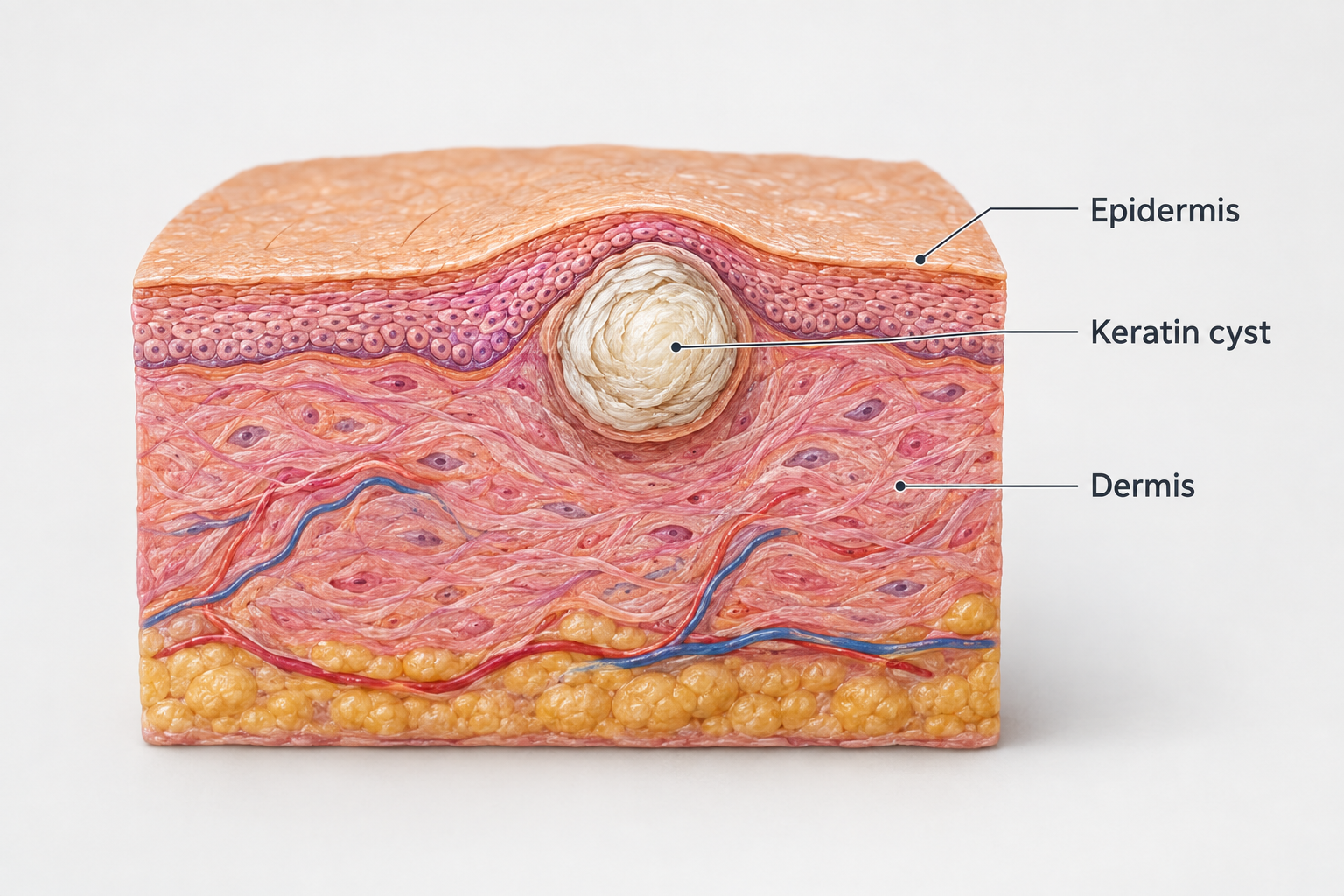

Milia under the eye are small, firm, white or yellowish bumps that sit just beneath the skin surface and can be surprisingly persistent. Unlike spots or whiteheads, they do not respond to squeezing and rarely disappear on their own in adults. This guide explains what they are, why they form in the delicate periocular area, which removal options are available, and how to reduce the risk of them returning.
Key Takeaways
- Milia are benign keratin-filled cysts trapped beneath the skin surface — they are harmless but tend to persist in adults without treatment.
- The thin skin around the eyes is particularly prone to milia because it has fewer oil glands and is often exposed to occlusive eye creams and cumulative sun damage.
- Professional removal by a trained practitioner is the safest and most reliable approach; at-home extraction carries a real risk of scarring and infection in this sensitive area.
- Topical retinoids and alpha hydroxy acids can support cell turnover and help prevent new milia, but must be used carefully near the eye area.
- Choosing non-comedogenic eye products and applying daily SPF are the most practical long-term prevention steps.
What Are Milia Under the Eyes?
Milia — sometimes called milk spots — are tiny keratin-filled cysts that form when dead skin cells become trapped beneath the skin surface rather than shedding normally. Each milium is essentially a small pocket of keratin protein enclosed by a thin cyst wall, sitting just below the epidermis. They typically measure one to two millimetres in diameter, appear white or pale yellow, and feel firm to the touch. Because they have no pore opening to the surface, they cannot be squeezed out like a conventional spot.
Milia are classified as either primary or secondary. Primary milia arise spontaneously without an obvious trigger and are the type most commonly seen under the eyes in adults. Secondary milia develop in response to skin trauma, injury, or certain medical treatments — for example, after a burn, a blister, aggressive dermabrasion, or prolonged corticosteroid use. Understanding which type is present can help guide the most appropriate management approach.
In newborns, milia are extremely common and typically resolve within a few weeks without any intervention. Adult under-eye milia behave quite differently: they are far less likely to self-resolve and may persist for months or years if left untreated. This distinction is worth understanding before deciding on a management plan.
Why Do Milia Form Around the Eyes Specifically?
The skin immediately around the eyes — the periocular area — is among the thinnest on the entire face, often measuring less than half a millimetre. This delicate periocular skin has fewer sebaceous (oil) glands than skin elsewhere, which means it produces less natural lubrication and relies more heavily on topical products for moisture. That reliance creates a specific vulnerability: heavy or occlusive eye creams can trap dead skin cells and keratin at the surface, preventing normal shedding and encouraging milia to form.
Cumulative sun damage is another significant contributing factor. UV exposure over time disrupts the orderly process of skin cell turnover, making it harder for the outermost skin cells to shed efficiently. The constant movement of blinking also subjects the eyelid skin to repeated mechanical stress, which may contribute to keratin becoming trapped beneath the surface.
Additional risk factors include oil-based or heavy makeup worn close to the eye, a genetic predisposition to slower cell turnover, underlying skin conditions such as rosacea or eczema, and — in the case of secondary milia — previous procedures or injuries to the area. Long-term use of topical corticosteroids around the eyes can thin the skin further and impair its natural renewal process, increasing the likelihood of secondary milia developing.
Milia vs. Whiteheads and Other Look-Alike Conditions
Milia are frequently confused with whiteheads, but the two conditions are meaningfully different. A whitehead (closed comedone) is a blocked pore containing a mixture of sebum, dead skin cells, and sometimes bacteria; it has a pore opening and can, in principle, be expressed. A milium contains only keratin and has no pore opening — which is why attempting to squeeze one typically produces nothing and risks damaging the surrounding skin.
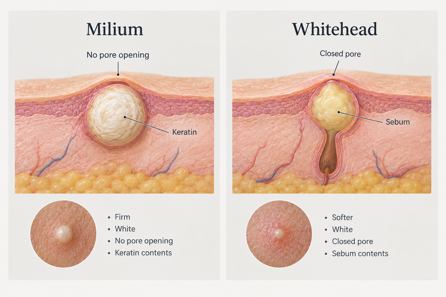
| Feature | Milia | Whitehead |
|---|---|---|
| Contents | Trapped keratin (protein) | Sebum and dead skin cells |
| Pore opening | None | Closed pore present |
| Texture | Firm, pearl-like | Softer, slightly compressible |
| Typical location | Under eyes, cheeks, nose | Forehead, chin, nose |
| Response to squeezing | Does not express | May express with pressure |
| Self-resolution (adults) | Unlikely without treatment | May resolve with skincare |
Other conditions that can resemble milia include sebaceous hyperplasia (enlarged oil glands that appear as yellowish, slightly indented bumps), flat warts, and — rarely — idiopathic calcinosis cutis. If there is any uncertainty about what a bump is, a dermatologist can confirm the diagnosis before any treatment is attempted.
Professional Removal Methods
Professional removal is the most reliable and safest route for under-eye milia. Because the skin in this area is so thin and close to the eye itself, precision matters considerably. Several techniques are used in clinical practice, and the most appropriate one depends on the number of milia, their depth, the patient's skin tone, and the practitioner's available equipment.
Manual Extraction (Deroofing)
Manual extraction — also called deroofing — is the most widely used method and is suitable for most patients. The practitioner cleanses the skin, then uses a sterile lancet or fine sterile needle to make a tiny incision in the cyst wall. The keratin plug is then gently lifted or expressed using a comedone extractor or light manual pressure. The procedure takes only a few seconds per milium and, when performed correctly by a trained professional, leaves minimal trauma to the surrounding tissue. Most patients experience only mild discomfort. A small red mark or pinpoint bleeding is normal immediately afterwards and typically settles within 24 to 48 hours.
Cryotherapy
Cryotherapy uses liquid nitrogen to freeze the milia, causing the cyst wall to break down so that the trapped keratin can be shed naturally as the skin heals. It is a quick in-clinic procedure but can cause temporary redness, blistering, and — particularly in individuals with skin of colour — a risk of post-inflammatory hyperpigmentation. For this reason, cryotherapy is generally used more cautiously in darker skin tones, and a thorough discussion of the hyperpigmentation risk is important before proceeding.
Laser Ablation
Laser ablation uses focused light energy to vaporise the milia with a high degree of precision. Erbium YAG and fractional CO2 lasers are the most commonly used devices. Erbium YAG laser is generally preferred over CO2 or Nd:YAG lasers for patients with skin of colour because it carries a lower hyperpigmentation risk — a nuance that is important to discuss with your practitioner if this applies to you. Laser treatment is particularly useful for multiple or deeply seated milia, though it typically involves a slightly longer recovery period of five to seven days compared with manual extraction.
Electrodesiccation (Diathermy)
Electrodesiccation, sometimes called diathermy, uses a fine electrical current to destroy the milia. A very fine probe is applied to each cyst, delivering a small burst of energy that desiccates the keratin contents. It is effective but requires careful technique near the eye area, and the risk of scarring or pigmentation change is slightly higher than with manual extraction if not performed precisely.
Chemical Peels and Dermabrasion
Superficial chemical peels using glycolic acid or other alpha hydroxy acids, and controlled dermabrasion or microdermabrasion, can help address multiple milia across a wider area by accelerating cell turnover and thinning the outermost skin layer. These approaches are more commonly used as preventive or maintenance treatments rather than for targeted removal of individual milia, and they are generally not applied directly to the eyelid skin itself due to the sensitivity of the area.
What Happens During a Professional Removal Appointment?
A typical milia removal appointment begins with a consultation to confirm the diagnosis and discuss the most appropriate technique. The skin is cleansed and, depending on the method, a topical numbing cream may be applied — though many patients find manual extraction tolerable without anaesthesia given the small size of each milia. The practitioner works through each milium individually, which means the appointment length depends on how many are being treated. A session addressing a small cluster of milia might take 15 to 30 minutes in total.
Immediately after treatment, the treated area may appear slightly red or have tiny marks where the skin was incised. Most patients can return to normal activities the same day, though the skin should be treated gently for the following 48 hours. Full recovery from manual extraction is typically complete within three to five days. Laser-treated areas may take five to seven days to heal fully, with a small crust or scab forming over each treated spot before it falls away naturally.
Post-Treatment Aftercare Timeline
Following professional milia removal, a structured aftercare approach helps the skin heal cleanly and reduces the risk of complications such as infection or pigmentation changes.
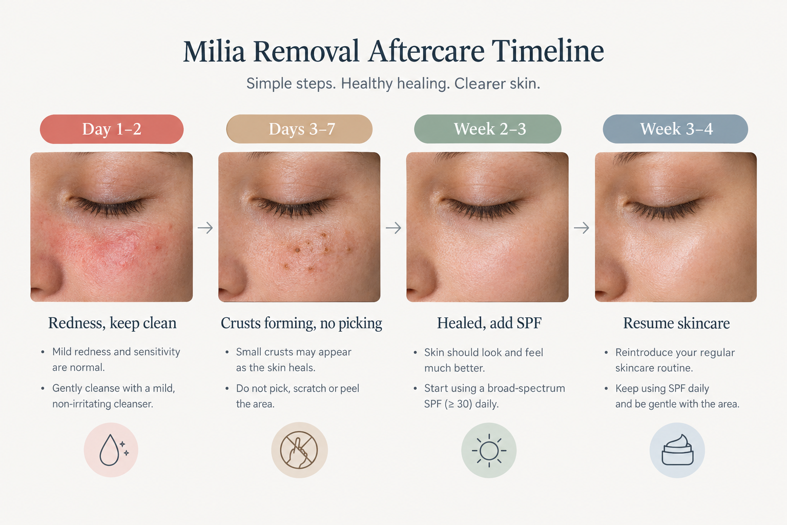
- Day 1–2: Expect mild redness, possible pinpoint bleeding, and slight swelling. Keep the area clean and dry. Avoid makeup, active skincare ingredients, and touching the treated skin. Apply a gentle, fragrance-free moisturiser if recommended by your practitioner.
- Days 3–5 (extraction) or Days 3–7 (laser): Small crusts or scabs may form over treated spots. Do not pick or remove them — allow them to fall away naturally. Continue to avoid exfoliants, retinoids, and AHAs on the treated area.
- Week 2–3: The skin should appear largely healed. Introduce SPF daily if you have not already done so. Avoid steam rooms, saunas, and intense exercise that causes heavy sweating until the skin is fully settled.
- Week 3–4 onwards: Resume normal skincare gradually. Discuss with your practitioner when it is appropriate to reintroduce retinoids or exfoliating acids as part of a prevention strategy.
Daily sun protection is particularly important after any milia removal procedure. UV exposure can worsen post-inflammatory hyperpigmentation, especially in individuals with medium to darker skin tones, and can also contribute to the formation of new milia over time.
At-Home Management: What Is Realistic?
At-home approaches cannot remove existing milia in the way that professional extraction can, but they can support cell turnover and may help prevent new ones from forming. The key distinction is between attempting to extract milia at home — which carries a genuine risk of scarring, infection, and hyperpigmentation in the thin under-eye skin — and using topical ingredients to gradually encourage the skin to shed more efficiently.
Topical Retinoids
Topical retinoids — including over-the-counter retinol and prescription-strength tretinoin or adapalene — are among the most evidence-supported options for improving skin cell turnover and may help prevent new milia from forming over time. However, an important nuance is often overlooked: retinoids and AHAs should generally not be applied directly onto the eyelid or the immediate under-eye skin, as this area is too thin and sensitive and the risk of irritation is high. Instead, these ingredients are typically applied to the surrounding cheek and orbital bone area, allowing a small amount of product migration to reach the periocular zone without direct application. If you are considering retinoids for milia prevention, discuss the appropriate application technique with a dermatologist or skincare professional.
Realistic expectations are also important here. Retinoids work gradually by accelerating cell turnover — they are unlikely to resolve established milia quickly, and consistent use over several months is typically needed before a meaningful preventive effect is seen on new milia formation.
Alpha Hydroxy Acids and Other Exfoliants
Alpha hydroxy acids (AHAs) such as glycolic acid and lactic acid, and beta hydroxy acids such as salicylic acid, can help loosen the bonds between dead skin cells and encourage more efficient shedding. Like retinoids, these should be used on the surrounding skin rather than directly on the eyelid, and formulations designed for the eye area should be chosen carefully. Polyhydroxy acids (PHAs) are a gentler alternative that may be better tolerated by sensitive skin types.
Gentle Cleansing and Warm Compresses
Regular gentle cleansing removes surface debris and excess product residue that can contribute to keratin becoming trapped. A warm, damp washcloth applied to the area for a few minutes can soften the skin and may support the natural shedding process, though it will not dissolve existing milia. These measures are most useful as part of a consistent preventive routine rather than as a treatment for established cysts.
Choosing Eye Products That Do Not Trigger Milia
The choice of eye cream and makeup products plays a meaningful role in both preventing and managing milia. Occlusive eye creams — those containing petrolatum, mineral oil, or heavy silicones — can create a barrier that traps dead skin cells and keratin beneath the surface, particularly in individuals already prone to milia. Switching to lighter formulations is one of the most practical preventive steps available.
When selecting an eye product, look for non-comedogenic formulations that contain ingredients such as hyaluronic acid for hydration, niacinamide for barrier support, or lightweight peptides. Avoid products with petrolatum or mineral oil as primary ingredients if you are prone to milia. Similarly, oil-based makeup removers used around the eye area should be rinsed away thoroughly rather than left on the skin.
When to See a Dermatologist or Specialist
Most under-eye milia can be assessed and treated by a trained aesthetic practitioner or dermatologist. However, milia that sit very close to the eyelid margin — the edge of the eyelid itself — may require assessment by an oculoplastic surgeon or ophthalmologist, as treatment in this precise location carries additional considerations related to eye safety. If you are unsure whether what you have is milia or another condition, a dermatologist can confirm the diagnosis and recommend the most appropriate management.
It is also worth seeking professional advice if milia appear suddenly in large numbers, if they are associated with skin fragility or blistering, or if they develop after a new medication or skincare product — as these patterns may suggest secondary milia with an underlying cause that warrants attention.
Cost and Practical Considerations
Milia removal is considered a cosmetic procedure and is not typically covered by health insurance. The cost varies depending on the technique used, the number of milia being treated, the type of clinic or specialist, and geographic location. Manual extraction of a small number of milia is generally the most affordable option. Laser ablation tends to cost more per session but may be more efficient for larger numbers of milia or for patients in whom manual extraction is less suitable.
Multiple sessions may occasionally be needed, particularly if new milia continue to form. A consultation fee is usually charged separately and is worth factoring into the overall cost. Asking for a clear breakdown of fees before committing to treatment is always reasonable.
Frequently Asked Questions
Can milia under the eyes go away on their own?
In adults, under-eye milia rarely resolve without some form of intervention. Unlike milia in newborns — which typically clear within a few weeks — adult milia tend to persist indefinitely because the keratin-filled cyst has no natural route to the skin surface. Professional removal or a consistent retinoid-based skincare routine offers the most reliable path to resolution.
Is it safe to try to remove milia at home?
Attempting to extract milia at home using a needle or pin is not recommended, particularly in the under-eye area. The skin here is extremely thin, and an imprecise incision risks scarring, infection, and hyperpigmentation. At-home management is best limited to supportive skincare — gentle exfoliation, appropriate topical ingredients, and non-comedogenic products — rather than extraction.
How many professional sessions will I need?
For a defined cluster of existing milia, a single professional extraction session is often sufficient. However, if new milia continue to form due to ongoing triggers — such as an occlusive eye cream or cumulative sun damage — further sessions may be needed over time. Addressing the underlying cause is as important as removing the existing cysts.
Does milia removal hurt?
Manual extraction using a sterile lancet causes minimal discomfort for most patients — typically described as a brief, sharp sensation. Laser ablation may involve slightly more discomfort, and a topical numbing cream is often applied beforehand. The under-eye area is sensitive, so some degree of awareness during treatment is normal, but the procedures are generally well tolerated.
Can retinol get rid of existing milia?
Retinol and other topical retinoids work by accelerating cell turnover, which may gradually help the skin shed trapped keratin over time. However, they are unlikely to resolve established milia quickly — consistent use over several months is typically needed, and results vary between individuals. Retinoids are more reliably useful as a preventive measure than as a treatment for existing cysts.
Are there any risks specific to darker skin tones?
Yes. Cryotherapy and certain laser treatments carry a higher risk of post-inflammatory hyperpigmentation in individuals with skin of colour. For darker skin tones, Erbium YAG laser is generally preferred over CO2 or Nd:YAG lasers because it has a lower hyperpigmentation risk. Manual extraction is often the safest first-line option across all skin tones when performed by an experienced practitioner. Discussing your skin tone and pigmentation history with your practitioner before any procedure is important.
Making an Informed Decision About Milia Removal
Under-eye milia are benign and pose no medical risk, but their persistence and visibility make them a reasonable concern to address. Professional removal — particularly manual deroofing with a sterile lancet — is safe, effective, and involves minimal downtime when carried out by a trained practitioner. Laser ablation offers a useful alternative for multiple or deeper milia, with the choice of laser device mattering particularly for individuals with skin of colour.
At-home approaches have a meaningful role in prevention but are not a substitute for professional extraction of established milia. Switching to non-comedogenic eye products, applying SPF daily, and introducing retinoids carefully to the surrounding skin area are the most practical steps for reducing the likelihood of new milia forming after treatment.
If you are considering milia removal, a consultation with a dermatologist or experienced aesthetic practitioner is the appropriate starting point. They can confirm the diagnosis, assess whether any underlying triggers need addressing, and recommend the technique most suited to your skin type, the number of milia present, and your individual circumstances.
