
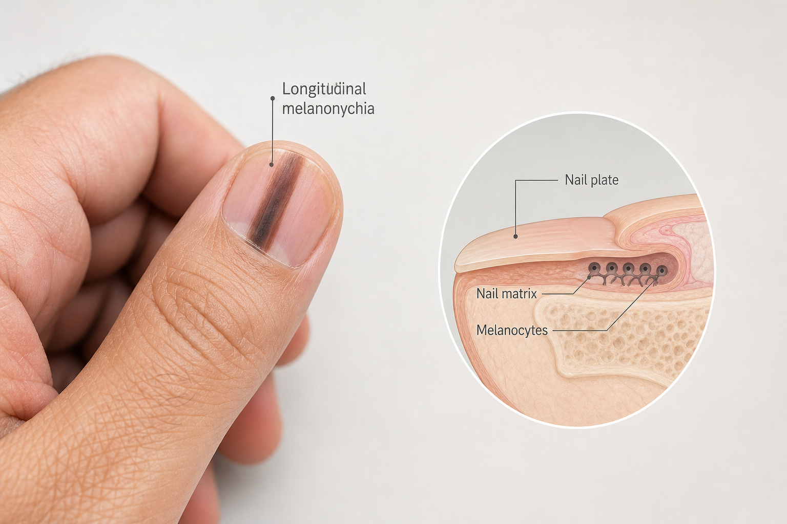

A black line on the nail — medically known as melanonychia — is one of the most anxiety-provoking things a person can notice on their own body. In most cases it is entirely harmless, but in a small number of cases it can be an early sign of subungual melanoma, a form of skin cancer that develops beneath the nail. Understanding the difference between a benign pigmented band and a warning sign that requires urgent evaluation is genuinely important, and this article explains both clearly.
Key Takeaways
- A black or dark stripe running along the nail is called longitudinal melanonychia and has many possible causes, most of which are benign.
- Racial or ethnic pigmentation is the most common cause in people with darker skin tones, affecting the majority of African American adults by age 50.
- Subungual melanoma is rare but serious; certain features — including band width, irregular borders, and Hutchinson’s sign — help distinguish it from benign causes.
- A dermatologist can use dermoscopy and, where necessary, nail matrix biopsy to reach an accurate diagnosis; self-diagnosis is not reliable.
- Any black line on the nail that is new, changing, or accompanied by nail destruction should be assessed by a qualified clinician promptly.
What Is a Black Line on the Nail?
The nail plate — the hard structure you see and trim — is produced by the nail matrix, a layer of specialized cells beneath the base of the nail. Within the nail matrix sit melanocytes, the same pigment-producing cells found in skin. When these melanocytes become active or increase in number, they deposit melanin into the growing nail plate, creating a visible dark stripe that travels from the base of the nail toward the tip. This stripe is called longitudinal melanonychia, and it can appear brown, dark brown, grey, or black depending on the depth of pigment deposition and the individual’s skin tone.
Melanonychia can affect a single nail (monodactylous) or multiple nails simultaneously (polydactylous). It can also be classified by pattern: longitudinal melanonychia runs from the cuticle to the free edge and is by far the most common form; transverse melanonychia crosses the nail horizontally; and diffuse melanonychia involves the entire nail plate. The longitudinal form is the one most likely to prompt concern and the one most thoroughly studied in relation to nail melanoma.
Benign Causes of a Dark Stripe on the Nail
The overwhelming majority of pigmented nail bands have a benign explanation. Understanding the most common causes helps put the finding in context, though it does not replace professional evaluation.
Racial and Ethnic Pigmentation
Racial melanonychia — also called ethnic pigmentation or physiological melanonychia — is the single most common cause of longitudinal melanonychia worldwide. It results from melanocytic activation rather than an abnormal increase in melanocyte numbers. Prevalence rises sharply with Fitzpatrick skin type: studies estimate that up to 77–100% of African American adults develop at least one pigmented nail band by the age of 50, compared with approximately 10–20% of Japanese adults and roughly 1% of white adults. The bands are typically well-defined, uniform in colour, and stable over time. Multiple nails are often involved. This pattern is considered a normal variant and does not require treatment.
Trauma and Mechanical Causes
Repetitive friction, nail biting, tight footwear, or a single significant impact can stimulate melanin production in the nail matrix. Trauma-related melanonychia tends to affect the nails most exposed to pressure — commonly the great toenail or the thumbnail — and may resolve once the source of friction is removed. A subungual hematoma (bruise under the nail) caused by a direct blow looks different: it typically appears as a dark, often purplish-red patch rather than a defined stripe, and it migrates toward the nail tip as the nail grows out over weeks to months.
Nail Matrix Nevus
A nail matrix nevus is a benign cluster of melanocytes within the nail matrix, analogous to a mole on the skin. It is the most common cause of longitudinal melanonychia in children and is also seen in adults. The band produced by a nail matrix nevus is usually a single, well-demarcated stripe with relatively uniform colour. In children, nail matrix nevi are almost always benign, and subungual melanoma in paediatric patients is extremely rare. In adults, a new or changing nevus warrants closer clinical scrutiny.
Systemic Diseases and Nutritional Deficiencies
Several systemic conditions can trigger melanocytic activation across multiple nails simultaneously. Addison’s disease (adrenal insufficiency) causes elevated levels of adrenocorticotropic hormone, which stimulates melanocytes throughout the body including the nail matrix, often producing diffuse or multi-nail pigmentation. Cushing’s syndrome, HIV infection, and certain connective tissue disorders have also been associated with nail pigmentation. Nutritional deficiencies — particularly vitamin B12 deficiency — are a recognised but underappreciated cause, especially in individuals following plant-based diets or those with malabsorption conditions. When multiple nails are affected and no other explanation is apparent, a clinician may investigate systemic causes.
Drug-Induced Melanonychia
A significant number of medications can cause drug-induced melanonychia by stimulating melanin production in the nail matrix. The effect is usually reversible once the medication is stopped, though this can take many months given the slow growth rate of nails. The table below lists the most commonly implicated drug categories and examples.
| Drug Category | Examples |
|---|---|
| Chemotherapy agents | Cyclophosphamide, doxorubicin, bleomycin, hydroxyurea |
| Antimalarials | Chloroquine, hydroxychloroquine |
| Antibiotics | Minocycline, zidovudine (AZT) |
| Anticonvulsants | Phenytoin |
| Biologics | Infliximab |
| Heavy metals | Arsenic, thallium, mercury (toxic exposure) |
If you have recently started a new medication and notice new nail pigmentation across several nails, mention this to your prescribing clinician. Do not stop prescribed medication without medical advice.
Fungal Infection
Certain fungal organisms, particularly Candida species, can produce pigmented compounds within the nail plate that mimic longitudinal melanonychia. Fungal melanonychia is an underrecognised cause that can closely resemble a benign pigmented band or, in some cases, even subungual melanoma on visual inspection alone. A dermatologist may perform a potassium hydroxide (KOH) preparation or fungal culture to rule out infection when the clinical picture is ambiguous. Antifungal treatment resolves the pigmentation in confirmed cases.
When a Black Line on the Nail May Be Serious
Subungual melanoma — also called nail unit melanoma (NUM) or nail apparatus melanoma (NAM) — is a rare but potentially life-threatening malignancy that arises from melanocytes within the nail unit. It accounts for approximately 0.7–3.5% of all melanomas in white populations but a disproportionately higher percentage in people of African, Asian, and Hispanic descent, where it may represent 15–35% of melanoma cases. Because it is often mistaken for a benign pigmented band, bruise, or fungal infection, diagnosis is frequently delayed — and delayed diagnosis is associated with a significantly worse prognosis compared with cutaneous melanoma detected at an equivalent stage.
The ABCDEF Rule for Nail Melanoma
The ABCDEF rule, developed by Levit and colleagues, provides a structured framework for identifying features that raise concern for subungual melanoma. It is a useful clinical guide, but it is important to acknowledge its limitations: research has shown that the ABCDEF criteria cannot reliably distinguish benign longitudinal melanonychia from subungual melanoma in all cases, and dermoscopic assessment by a trained clinician is more accurate than visual inspection alone. The rule should prompt professional evaluation, not serve as a substitute for it.
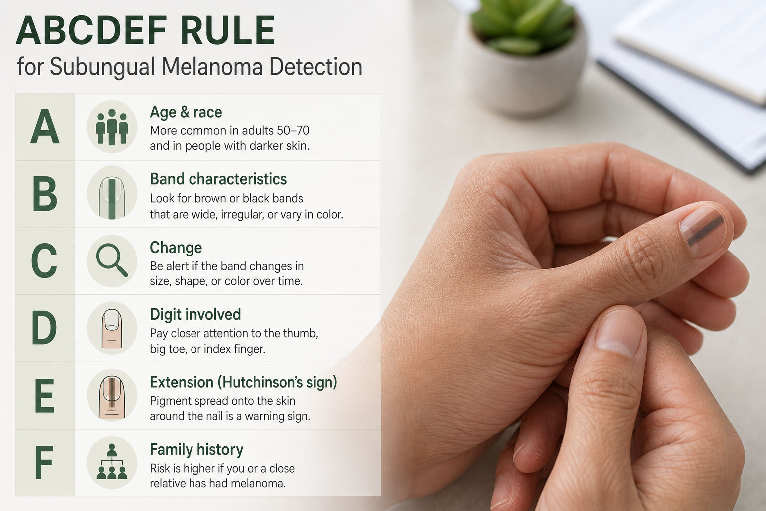
| Letter | Feature | What to Look For |
|---|---|---|
| A | Age and race | Peak incidence age 40–70; higher risk in African American, Asian, Hispanic individuals; also the most common nail malignancy in these groups |
| B | Band characteristics | Brown or black band; width ≥3 mm; blurred or irregular borders; multiple shades within the band |
| C | Change | Rapid increase in size, width, or colour change despite treatment |
| D | Digit involved | Thumb, index finger, or great toe most commonly affected; single digit involvement |
| E | Extension | Hutchinson’s sign — pigment extending onto the surrounding skin of the nail fold or cuticle |
| F | Family or personal history | Personal or family history of melanoma or dysplastic naevus syndrome |
Hutchinson’s Sign and the Pseudo-Hutchinson Sign
Hutchinson’s sign refers to the extension of dark pigmentation from beneath the nail onto the surrounding periungual skin — the cuticle, nail folds, or fingertip. When genuine, it is a significant warning sign for subungual melanoma and should prompt urgent dermatological assessment. However, a clinically important distinction exists: the pseudo-Hutchinson sign describes a situation where pigment from a benign nail band is visible through the translucent cuticle, giving the false impression that pigment has spread onto the skin surface. The pseudo-Hutchinson sign is associated with benign causes including racial melanonychia and nail matrix nevi, and it does not carry the same clinical significance. Distinguishing between the two requires dermoscopy and clinical expertise — it cannot be done reliably by visual inspection alone, which is one reason self-assessment has clear limits.
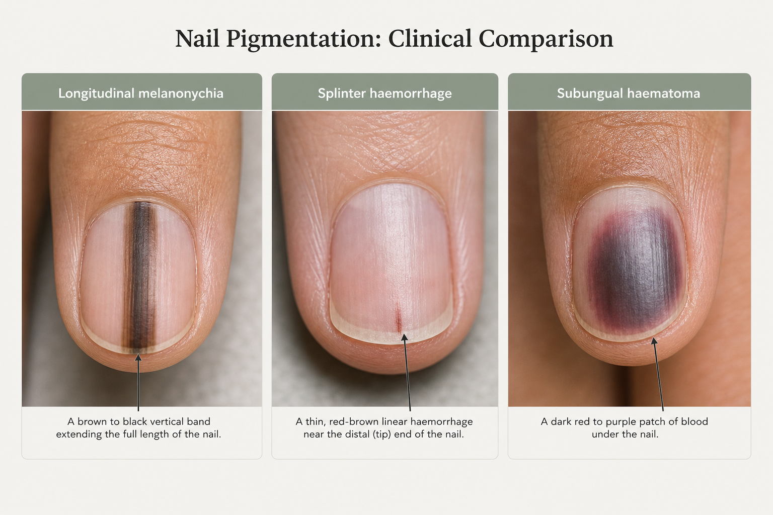
Melanonychia vs. Splinter Haemorrhage vs. Subungual Bruise
Not every dark mark under the nail is melanonychia. The table below compares the three most commonly confused presentations to help readers understand what they may be looking at — while emphasising that professional assessment remains the only reliable way to reach a diagnosis.
| Feature | Longitudinal Melanonychia | Splinter Haemorrhage | Subungual Haematoma (Bruise) |
|---|---|---|---|
| Appearance | Brown, dark brown, or black stripe running full length of nail | Thin dark red or brown vertical line, often near the nail tip | Dark red, purple, or black patch; may cover large area of nail |
| Cause | Melanin deposition from nail matrix melanocytes | Tiny blood vessel rupture; associated with trauma or systemic disease (e.g. endocarditis) | Direct trauma causing bleeding beneath the nail plate |
| Behaviour over time | Remains in fixed position relative to nail base; does not migrate | Migrates toward nail tip as nail grows; fades over weeks | Migrates toward nail tip as nail grows; colour changes from dark to yellow-brown |
| Number of nails | One or multiple | Usually one; multiple may suggest systemic disease | Usually one (the injured nail) |
| Action needed | Dermatological assessment if new, changing, or concerning features present | Single: monitor; multiple without trauma: medical review | Monitor; seek review if very painful or nail is at risk of loss |
A splinter haemorrhage is a thin, dark red or brown vertical line caused by rupture of a small blood vessel beneath the nail. A single splinter haemorrhage after minor trauma is common and harmless. Multiple splinter haemorrhages appearing without obvious trauma can occasionally be associated with systemic conditions including infective endocarditis and should prompt medical review. Unlike melanonychia, both splinter haemorrhages and subungual haematomas migrate toward the nail tip as the nail grows — a useful distinguishing feature. Fingernails grow approximately 3 mm per month, meaning a full nail cycle takes roughly six months; toenails grow more slowly, taking 12–18 months to fully replace. A dark mark that moves with nail growth is almost certainly blood-related rather than pigment-related.
How Is Melanonychia Diagnosed?
A dermatologist evaluating a pigmented nail band will typically follow a structured process. Understanding what to expect at the appointment can reduce anxiety and help you prepare.
Clinical History and Visual Examination
The clinician will ask about the duration of the band, whether it has changed in width, colour, or borders, any relevant medications, personal or family history of melanoma, and whether trauma to the nail has occurred. A thorough visual examination of all nails, the surrounding skin, and the rest of the body follows. The clinician will assess band width, colour uniformity, border regularity, and whether Hutchinson’s sign is present.
Dermoscopy and Onychoscopy
Dermoscopy — also called onychoscopy when applied specifically to the nail — uses a handheld magnifying device with polarised light to examine the nail plate and periungual skin in detail. Dermoscopy significantly improves the ability to distinguish benign from potentially malignant pigmented bands compared with the naked eye alone. Features assessed include the regularity of longitudinal lines within the band, colour variation, the presence of dots or globules, and the appearance of the cuticle. Dermoscopy is non-invasive and causes no discomfort.
Nail Matrix Biopsy
When dermoscopy and clinical assessment cannot confidently exclude malignancy, a nail matrix biopsy is performed. This involves taking a small tissue sample from the nail matrix under local anaesthetic for histological examination. Nail matrix biopsy is the definitive diagnostic test for subungual melanoma. It is a minor surgical procedure that may cause temporary nail deformity, and the decision to perform it is made carefully based on the overall clinical picture. A KOH preparation or fungal culture may also be requested if fungal melanonychia is suspected.
Red Flags: When to See a Dermatologist Promptly
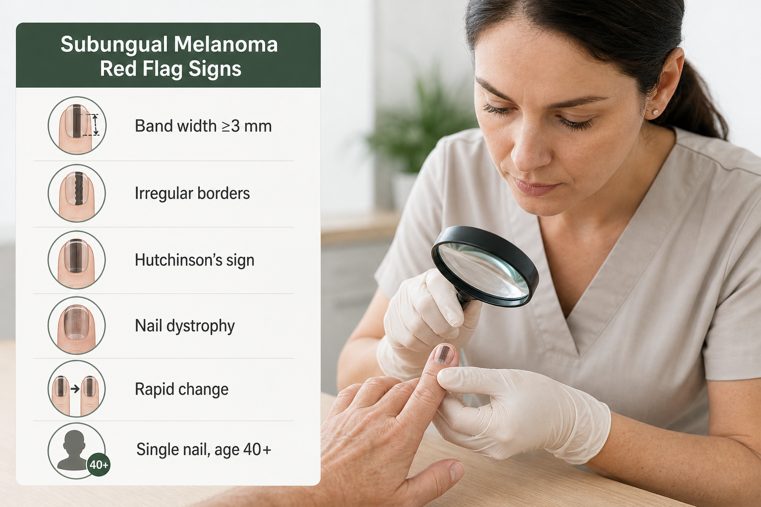
The following features should prompt timely professional assessment rather than a wait-and-see approach. This list is not exhaustive, and any new or changing nail pigmentation that concerns you is a valid reason to seek evaluation.
- A pigmented band that is new and has appeared without obvious cause
- A band that is widening, darkening, or changing in any way
- Band width of 3 mm or greater
- Irregular, blurred, or poorly defined borders within the band
- Multiple shades of brown or black within a single band
- Pigment extending onto the cuticle or surrounding skin (possible Hutchinson’s sign)
- Nail dystrophy — splitting, thinning, or destruction of the nail plate
- A single nail affected in a person over 40 with no obvious benign explanation
- Personal or family history of melanoma
- A band that has not resolved or migrated after several months (ruling out a haematoma)
Melanonychia in Children
Longitudinal melanonychia in children is almost always benign. The most common cause in paediatric patients is a nail matrix nevus, followed by racial pigmentation in children with darker skin tones. Subungual melanoma is exceptionally rare in children and adolescents. Despite this reassuring picture, a new pigmented band in a child should still be assessed by a dermatologist, particularly if it is wide, has irregular features, or is changing rapidly. Dermoscopy is well-tolerated in children and can usually provide sufficient information to guide management without the need for biopsy.
Treatment and Management Options
Management of melanonychia depends entirely on the underlying cause and whether the band is considered benign or potentially malignant.
Benign Melanonychia: Watch, Wait, and Address the Cause
When a pigmented band is confidently identified as benign — for example, racial melanonychia or a stable nail matrix nevus — no medical treatment is required. The clinician may recommend photographic documentation and periodic review to monitor for any change. If a medication is responsible, the prescribing clinician may consider an alternative where clinically appropriate. Nutritional deficiencies should be corrected. Fungal melanonychia is treated with antifungal medication, and the pigmentation typically resolves as the nail grows out.
Cosmetic Management
For individuals who are bothered by the appearance of a benign pigmented band, options include nail polish or cosmetic camouflage. Laser therapy has been explored as a cosmetic treatment for benign melanonychia, particularly in cases of racial pigmentation, but evidence for its effectiveness and safety in this context remains limited, and there is a theoretical risk of masking a change in the band if monitoring is not maintained. Any cosmetic approach should only be considered after a benign diagnosis has been confirmed.
Subungual Melanoma: Surgical Treatment
Confirmed subungual melanoma requires surgical treatment. Historically, amputation of the affected digit was standard practice, but more conservative surgical approaches — including wide local excision of the nail unit — are now used in appropriate cases depending on the stage and location of the tumour. Early-stage nail unit melanoma has a significantly better prognosis than advanced disease, reinforcing the importance of prompt diagnosis. Subungual melanoma generally carries a poorer prognosis than cutaneous melanoma detected at a comparable stage, largely because diagnosis is frequently delayed by months or years. Sentinel lymph node biopsy and systemic staging investigations may be required depending on tumour thickness.
Frequently Asked Questions
Is a black line on the nail always cancer?
No. The vast majority of pigmented nail bands are benign. Common causes include racial pigmentation, trauma, nail matrix nevi, medications, and systemic conditions. Subungual melanoma is rare, but because it can present as a dark stripe, any new or changing band should be assessed by a dermatologist to rule it out.
Can a black line on the nail go away on its own?
It depends on the cause. A subungual haematoma (bruise) will migrate toward the nail tip and disappear as the nail grows out over several months. Drug-induced melanonychia may fade after the causative medication is stopped, though this can take six months or longer. Racial melanonychia and nail matrix nevi are typically permanent. A band that is genuinely moving toward the nail tip is more likely to be blood-related than pigment-related.
What is the difference between Hutchinson’s sign and the pseudo-Hutchinson sign?
Hutchinson’s sign is the extension of dark pigment from beneath the nail onto the surrounding skin of the cuticle or nail fold, and it is a warning sign for subungual melanoma. The pseudo-Hutchinson sign occurs when pigment from a benign band is visible through the translucent cuticle, mimicking true extension. Distinguishing between the two requires dermoscopy and clinical expertise.
Should I be worried if I have a black line on my nail and I am of African or Asian descent?
Longitudinal melanonychia is far more common in people with darker skin tones and is usually a normal variant in this population. However, subungual melanoma also occurs more frequently in people of African, Asian, and Hispanic descent as a proportion of all melanoma cases. Any band that is new, wide, irregular, or changing should be assessed regardless of skin tone.
How long does it take for a nail to grow out completely?
Fingernails grow approximately 3 mm per month, so a full nail cycle takes roughly six months. Toenails grow more slowly and may take 12–18 months to fully replace. This timeline is clinically useful: a dark mark that visibly migrates toward the nail tip over this period is consistent with a haematoma rather than melanonychia.
Can a fungal nail infection cause a black line?
Yes. Certain fungal organisms, particularly Candida species, can produce pigmented compounds within the nail that create a dark stripe resembling longitudinal melanonychia. Fungal melanonychia can be difficult to distinguish from other causes on visual inspection alone. A dermatologist may perform a KOH preparation or fungal culture to confirm or exclude infection.
What happens if I ignore a black line on my nail?
If the band is genuinely benign, ignoring it carries no medical risk. However, because subungual melanoma can present identically to a benign band in its early stages, ignoring a new or changing stripe risks delayed diagnosis of a potentially serious condition. Subungual melanoma has a significantly better prognosis when detected early. Professional assessment is always the safer choice when there is any doubt.
When to Act on a Black Line on Your Nail
A black line on the nail is a finding that deserves attention but not automatic alarm. In the majority of people — particularly those with darker skin tones — it reflects normal melanocytic activity or a benign cause that requires no treatment. The challenge is that subungual melanoma, though rare, can look identical to a harmless pigmented band in its early stages, and it carries a worse prognosis when diagnosis is delayed. The practical approach is straightforward: if a pigmented band is new, changing, wide, irregularly bordered, or accompanied by any of the red flags described in this article, arrange a dermatological assessment promptly. Dermoscopy and, where necessary, nail matrix biopsy provide the diagnostic clarity that visual inspection alone cannot. For bands that have been stable for years in a person with known risk factors for racial melanonychia, periodic monitoring with photographic documentation is a reasonable approach — but this decision should be made with a qualified clinician rather than independently. Early assessment is always preferable to prolonged uncertainty.
