

Picking or scratching a mole — whether accidentally or out of habit — is more common than most people admit, and the immediate reaction is often a mix of pain, bleeding, and worry. The good news is that a single incident of picking a benign mole is unlikely to cause serious harm. The more important concern is understanding what the risks actually are, which ones are overstated, and which warning signs genuinely warrant a visit to a board-certified dermatologist.
Key Takeaways
- Picking a mole typically causes bleeding, inflammation, and an open wound — all of which usually heal within a week or two with basic wound care.
- Picking a mole does not cause skin cancer, but it can destroy surface tissue that a dermatologist would otherwise examine or send for pathology, creating a delayed diagnosis risk.
- Infection is a real risk whenever the skin barrier is broken; watch for redness spreading beyond the wound, swelling, pus, or fever.
- Moles can regrow after picking due to residual pigment cells left in the dermis, and this regrowth — known as a recurrent naevus — can look irregular enough to mimic a concerning lesion.
- Any mole that bleeds without trauma, itches persistently, or changes in appearance should be assessed by a dermatologist regardless of whether it has been picked.
What a Mole Actually Is
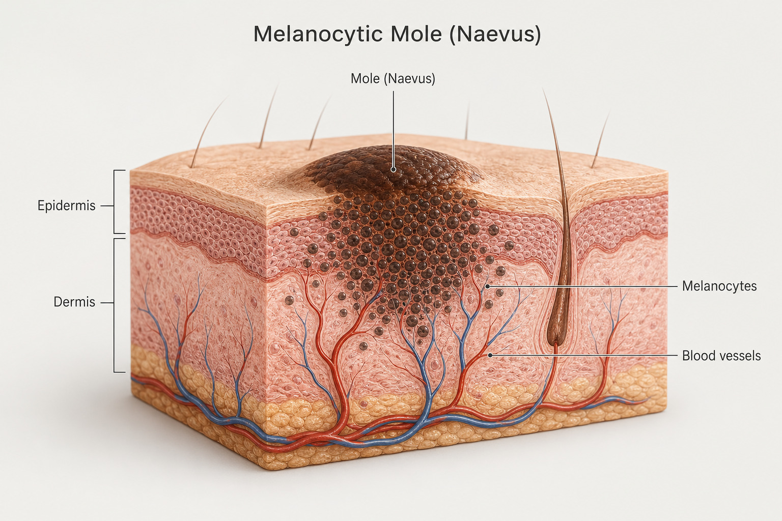
A mole — medically called a melanocytic naevus or nevus — is a cluster of melanocytes, the pigment-producing cells responsible for skin colour. These cells normally sit dispersed throughout the epidermis (the outer skin layer) and dermis (the deeper layer beneath). When melanocytes group together, they form the raised or flat pigmented spots most people recognise as moles. Most adults have between 10 and 40 moles, and the majority are entirely benign.
Moles fall into two broad categories. A congenital mole is present from birth and tends to be larger, sometimes covering a significant area of skin. An acquired mole develops during childhood or adulthood, usually as a result of sun exposure and genetic factors. Congenital moles carry a modestly higher lifetime risk of melanoma compared with acquired moles, which means picking or traumatising a congenital mole — and potentially obscuring its appearance — carries slightly higher stakes when it comes to monitoring.
Moles also contain small blood vessels that supply the melanocyte cluster with nutrients. This vascular supply is the reason even a minor scratch can produce a surprisingly brisk bleed.
What Happens Immediately When You Pick a Mole
The moment a mole is picked, scratched, or partially torn, several things happen in quick succession. The blood vessels within the mole are disrupted, causing bleeding that can seem disproportionate to the size of the lesion. The skin barrier is broken, exposing the underlying tissue to the environment. An inflammatory response begins almost immediately — the body sends increased blood flow and immune cells to the area, producing the familiar signs of redness, warmth, and swelling around the wound.
Pain or soreness is common and usually proportional to how deeply the mole was disturbed. A light scratch may cause only superficial discomfort, while pulling at a raised mole more forcefully can be genuinely painful and may remove a portion of the lesion entirely, leaving a raw open wound. Over the following hours, a scab typically forms as the wound begins to close. This scab is part of normal healing and should not be picked off, as doing so restarts the cycle of disruption and delays recovery.
Most uncomplicated cases heal within one to two weeks, provided the area is kept clean and left alone. Deeper wounds or repeated picking can extend healing time and increase the likelihood of permanent scarring.
Infection Risk: What to Watch For
An open wound on the skin is an entry point for bacteria. The hands carry a significant bacterial load, and any contact between unwashed fingers and a freshly picked mole introduces microorganisms directly into the disrupted tissue. Staphylococcus aureus is among the most common culprits in skin wound infections, though other bacteria present on the skin surface can also cause problems.
Signs that a picked mole has become infected include:
- Redness that spreads beyond the immediate wound edge — normal healing produces localised redness; spreading redness suggests bacterial involvement.
- Increasing swelling after the first 24–48 hours rather than gradual reduction.
- Pus or cloudy discharge from the wound.
- Warmth and tenderness that worsen rather than improve over several days.
- Fever or general malaise — these suggest the infection may be spreading beyond the local tissue and require prompt medical attention.
Basic wound care significantly reduces infection risk. After picking a mole, wash the area gently with mild soap and warm water, apply a thin layer of antibiotic ointment (available over the counter), and cover it with a clean adhesive bandage. Change the dressing daily and keep the area dry where possible. If signs of infection appear within three to five days, or if the wound shows no improvement after two weeks, see a doctor or dermatologist.
Does Picking a Mole Cause Scarring?
Scarring is a genuine and often underappreciated risk of picking moles. The depth of the wound largely determines the outcome. Superficial scratches that only disturb the epidermis tend to heal without a permanent mark. Deeper wounds that reach the dermis — particularly if the mole is raised and a portion is torn away — are more likely to leave a scar.
Repeated picking of the same mole compounds the risk considerably. Each cycle of disruption and healing lays down additional collagen in a less organised pattern than the original skin, gradually producing a thickened, irregular, or discoloured scar. In people prone to keloid or hypertrophic scarring, even a relatively minor wound can result in a raised, persistent scar that is cosmetically noticeable.
Scarring also has a practical clinical consequence: scar tissue can distort the appearance of the mole, making it harder for a dermatologist to assess whether any remaining pigmented cells look normal. This connects directly to the delayed diagnosis risk discussed in the next section.
Can Picking a Mole Cause Cancer?
This is the question most people are really asking, and the short answer is: picking a mole does not cause skin cancer. Cancer arises from genetic mutations within cells — primarily mutations in genes such as BRAF — that cause melanocytes to lose their normal growth regulation. Physical trauma to the skin surface does not directly trigger these mutations in otherwise healthy cells.
The more nuanced picture is that trauma to a mole that already contains abnormal or pre-cancerous cells may theoretically create an inflammatory wound environment that could influence cell behaviour, though this remains an area of ongoing research rather than established clinical fact. Some laboratory studies, including work using zebrafish models, have explored whether trauma combined with pre-existing preneoplastic cells might accelerate progression, but this has not been demonstrated to be a clinically significant risk from ordinary picking in humans.
What picking can do is remove or damage the surface tissue of a mole that might have been cancerous or atypical, meaning that the visible evidence is gone before a dermatologist has the chance to examine it. No tissue is sent for pathology, no biopsy is taken, and any abnormal cells that remain deeper in the skin are now hidden beneath a healing wound or scar. This is the delayed diagnosis risk — and it is arguably more important than the cancer-causation myth.
The Delayed Diagnosis Risk: Why This Matters More Than Most People Realise
When a dermatologist examines a suspicious mole, they are assessing its surface characteristics — colour variation, border irregularity, asymmetry, and size — alongside the patient’s history and, where indicated, dermoscopy (a magnified examination of the skin). If a mole has been partially picked away, the surface evidence that would inform that assessment is gone. The dermatologist is now looking at a healing wound or a scar rather than the original lesion.
The problem is compounded by the fact that cancerous or atypical cells may extend deeper into the dermis than the portion that was picked off. Those cells remain in place, but the surface marker that would have prompted investigation has been removed. A scar can also camouflage the irregular pigmentation or border changes that are characteristic of an atypical mole or early melanoma, potentially delaying recognition for months or years.
This is why dermatologists consistently advise against any form of home mole removal — not because picking causes cancer, but because it eliminates the opportunity for proper pathology. A shave excision or surgical excision performed by a trained clinician removes the mole in a controlled way, sends the tissue to a laboratory for analysis, and provides a definitive answer about whether the cells were normal. Picking a mole at home provides none of these safeguards.
Will a Mole Grow Back After Picking?
Yes, a mole can regrow after picking, and the regrowth may look different from the original lesion. When a mole is picked rather than professionally excised, residual pigment cells — melanocytes that were not removed — remain in the deeper layers of the dermis. These residual melanocytes can proliferate and repopulate the area, producing what is known as a recurrent naevus.
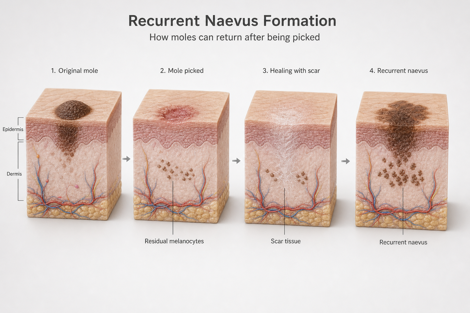
A recurrent naevus often appears within weeks to months of the original trauma. It may be lighter or darker than the original mole, and it frequently displays irregular pigmentation or borders that can look alarming. This irregular appearance is a direct result of the disorganised regrowth of melanocytes through scar tissue, rather than an indication that the mole has become cancerous. However, because a recurrent naevus can closely mimic the appearance of a melanoma, it should always be assessed by a dermatologist rather than monitored at home. The clinical history of prior trauma to the site is important information for the examining clinician.
Warning Signs: When a Mole Needs Professional Assessment
Whether or not a mole has been picked, certain characteristics should prompt a dermatology appointment. The ABCDE rule provides a practical framework for self-assessment:
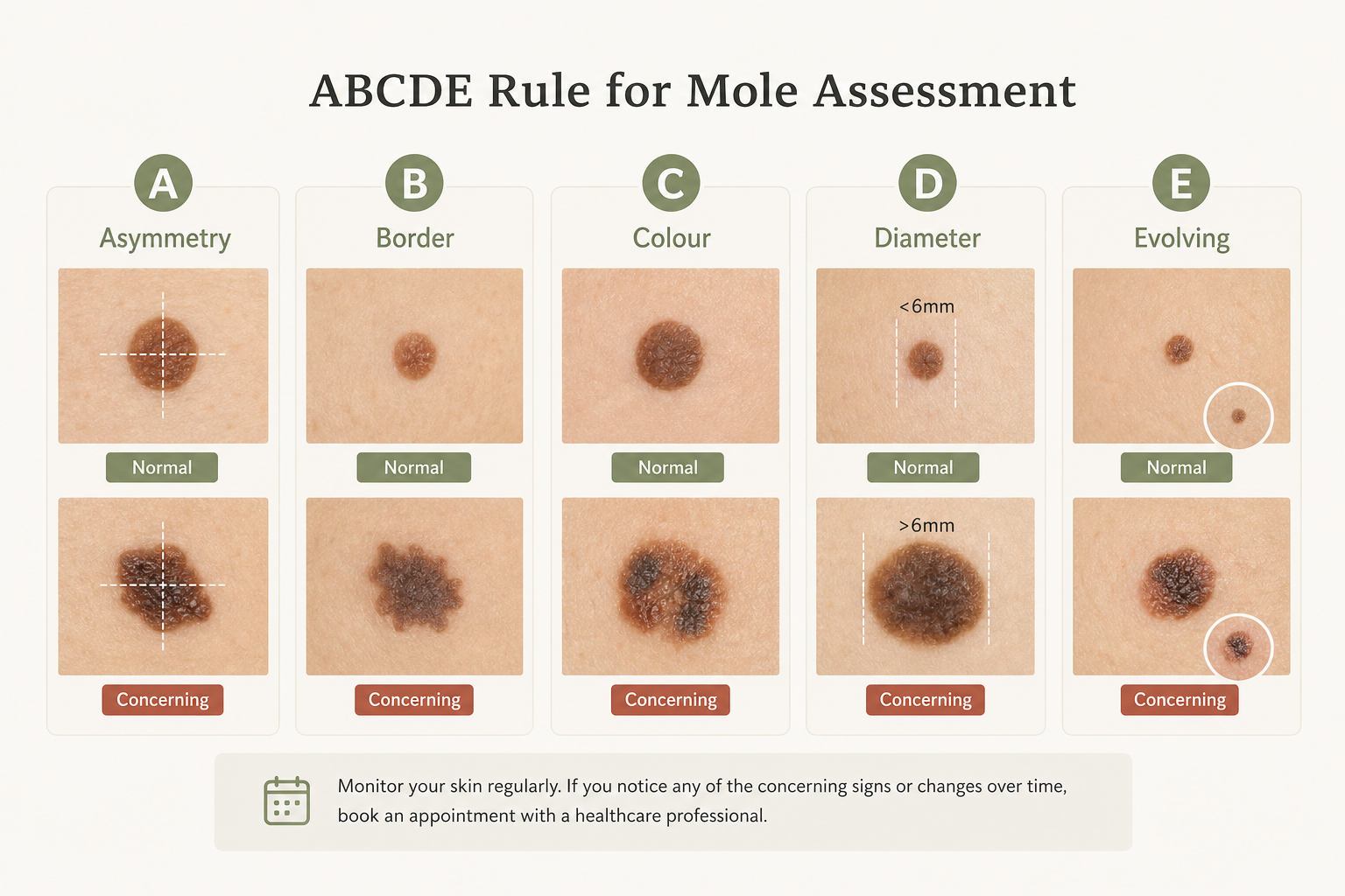
- Asymmetry — one half of the mole does not match the other.
- Border — the edges are irregular, ragged, notched, or blurred rather than smooth and well-defined.
- Colour — the mole contains multiple shades of brown, black, red, white, or blue rather than a uniform colour.
- Diameter — the mole is larger than approximately 6mm (roughly the size of a pencil eraser), though melanomas can be smaller.
- Evolving — the mole is changing in size, shape, colour, or texture, or has developed new symptoms such as bleeding, itching, or crusting.
On darker skin tones, melanoma warning signs can be subtler. Colour changes may be less immediately obvious, and lesions may appear on areas less commonly associated with sun exposure, such as the palms, soles, or under the nails. People with darker skin tones should be particularly attentive to the evolving criterion and to any new or changing lesion in these locations.
Beyond the ABCDE rule, any mole that bleeds without being scratched or knocked, itches persistently, or simply looks noticeably different from your other moles — sometimes called the “ugly duckling” sign — warrants professional evaluation.
First Aid Steps After Accidentally Picking a Mole
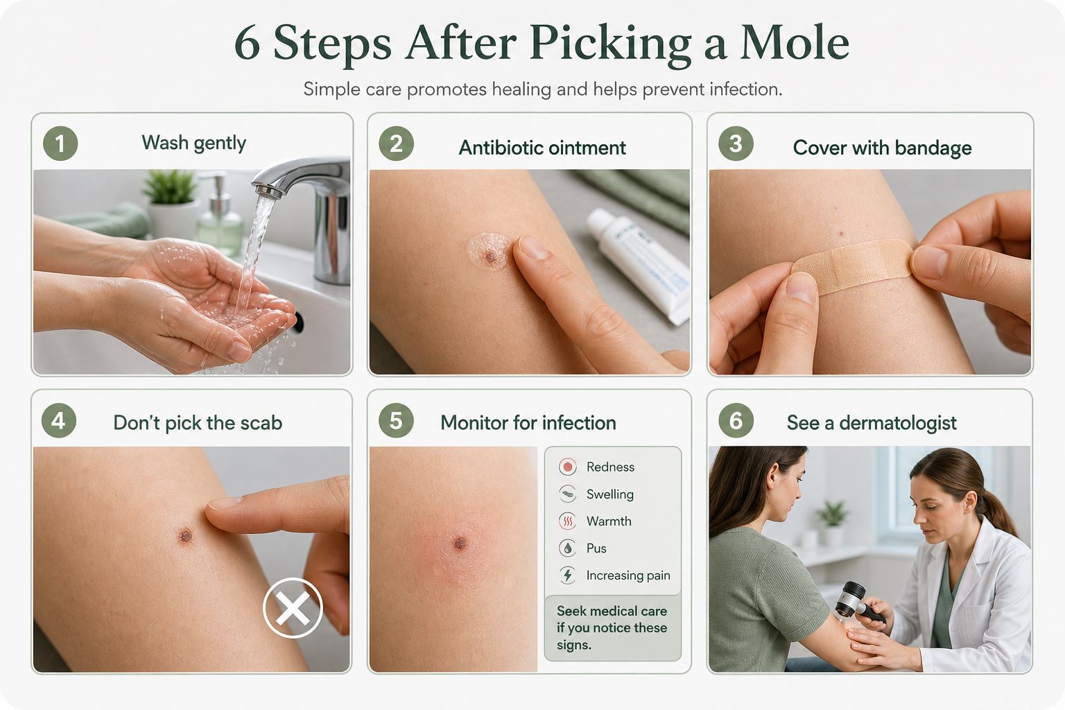
If you have already picked or scratched a mole, the following steps support safe healing and reduce the risk of complications:
- Wash the area gently with mild soap and warm water to remove bacteria and debris. Avoid scrubbing.
- Apply a thin layer of antibiotic ointment to the wound to reduce infection risk and support moist wound healing.
- Cover with a clean bandage and change it daily or whenever it becomes wet or dirty.
- Resist picking the scab that forms — removing it disrupts the healing process and increases scarring risk.
- Monitor for infection signs over the following three to five days: spreading redness, increasing swelling, pus, or fever.
- See a dermatologist if the wound does not heal within two weeks, if infection signs appear, if the mole was atypical before picking, or if you are unsure whether the mole had any concerning features.
Moles That Catch on Clothing or Jewellery
Raised moles — particularly those on the neck, underarms, bra line, or waistband area — are frequently caught on clothing, jewellery, or accessories. This type of accidental trauma is one of the most common reasons people find themselves dealing with a bleeding or partially disrupted mole. The same first aid principles apply, but prevention is straightforward: covering raised moles in friction-prone areas with a small adhesive bandage during activities that are likely to cause snagging, choosing clothing with smooth rather than rough seams in those areas, and removing jewellery that repeatedly catches on a mole can all reduce the frequency of accidental injury. If a particular mole is repeatedly traumatised by its location, professional removal is a sensible option to discuss with a dermatologist — both to prevent ongoing irritation and to obtain a pathology result.
At-Home Mole Removal Products: An Important Warning
A range of products marketed as at-home mole removal solutions — including mole removal pens that use electrical current, acid-based creams, freeze-off kits, and folk remedies such as apple cider vinegar or iodine — are widely available online. These products carry significant risks and are not a safe alternative to professional assessment.
The core problem with any at-home mole removal method is the same as with picking: no tissue is sent for pathology. A mole removal pen or acid cream destroys surface tissue without providing any information about whether the cells were normal. If the mole contained atypical or cancerous cells, those cells may remain in the deeper dermis while the surface evidence is gone — the same delayed diagnosis scenario described earlier. The US Food and Drug Administration (FDA) has issued warnings about unregulated mole removal devices and products, noting risks including chemical burns, permanent scarring, and the masking of potentially cancerous lesions. Professional removal by a board-certified dermatologist using shave excision, surgical excision, or punch biopsy provides both safe removal and a definitive pathology result.
Compulsive Mole Picking and Excoriation Disorder
For some people, picking at moles, scabs, or other skin irregularities is not an occasional accident but a repetitive, difficult-to-control behaviour. Excoriation disorder — also known as dermatillomania or compulsive skin picking — is a recognised body-focused repetitive behaviour that affects an estimated 1–5% of the population. It is associated with anxiety, obsessive-compulsive disorder (OCD), and other mental health conditions, and the picking often occurs automatically or in response to stress rather than as a deliberate choice.
If you find yourself repeatedly picking at moles or other skin areas despite wanting to stop, and if the behaviour is causing distress, skin damage, or social difficulty, this is worth discussing with a healthcare professional. Effective treatments exist, including cognitive behavioural therapy (CBT) with a habit reversal component, and addressing the underlying compulsive picking behaviour is important both for skin health and for overall wellbeing. A dermatologist can help manage the skin consequences while also directing you toward appropriate psychological support.
Professional Mole Removal: What It Involves
Professional mole removal is performed under local anaesthetic, meaning the area is numbed before any procedure begins. The method chosen depends on the mole’s size, depth, location, and whether there is any clinical concern about its appearance.
Shave excision involves using a fine blade to remove a raised mole flush with the skin surface. It is suitable for benign raised moles and produces a flat wound that heals relatively quickly. Surgical excision involves cutting out the mole and a small margin of surrounding skin, then closing the wound with sutures. This method is used when a mole is flat, when it has suspicious features, or when complete removal with clear margins is important. Punch biopsy uses a circular cutting tool to remove a small core of tissue and is often used for diagnostic purposes. In all cases, the removed tissue is sent to a pathology laboratory for analysis, providing a definitive assessment of the cells.
Laser removal is sometimes used for cosmetic removal of benign moles but does not provide a tissue sample for pathology, which is why it is generally not appropriate for moles with any atypical features. Cryotherapy (freezing) is similarly limited in this respect.
Frequently Asked Questions
Is it bad to pick a mole even if it looks completely normal?
Picking a mole that appears entirely normal is unlikely to cause serious harm, but it does carry risks of bleeding, infection, and scarring. It also removes surface tissue that a dermatologist might otherwise examine. The safest approach is to leave moles alone and have any that concern you assessed professionally.
What should I do if I accidentally scratched off a mole completely?
Clean the wound with mild soap and water, apply antibiotic ointment, and cover it with a bandage. Monitor for infection signs over the following days. See a dermatologist, particularly if the mole had any features that concerned you before the incident, or if the wound does not heal normally within two weeks.
Can a mole that has been picked look cancerous even if it is not?
Yes. A recurrent naevus — the regrowth that occurs when residual pigment cells repopulate a picked mole — can display irregular pigmentation and borders that closely resemble a melanoma. This is why any mole that looks different after a picking incident should be assessed by a dermatologist, who can take the history of trauma into account.
How long does a picked mole take to heal?
Most superficial wounds heal within one to two weeks with basic wound care. Deeper wounds, or those that become infected or are repeatedly disturbed, may take longer. Persistent non-healing after two weeks warrants medical review.
Should I be worried if a mole bleeds after being caught on clothing?
Bleeding from a mole that has been physically caught or scratched is usually a straightforward mechanical injury and not a cause for immediate alarm. Apply basic wound care and monitor healing. However, if the same mole bleeds repeatedly without obvious trauma, or if it has any features that concern you, arrange a dermatology assessment.
Is it safe to use a mole removal pen or cream at home?
No. At-home mole removal products including mole removal pens, acid creams, and freeze-off kits carry risks of chemical burns, scarring, and — most importantly — the destruction of tissue that should be sent for pathology. The FDA has warned against unregulated mole removal devices. Professional removal by a board-certified dermatologist is the only method that combines safe removal with a definitive pathology result.
What is the difference between a normal mole and an atypical mole?
An atypical mole (also called a dysplastic nevus) is a mole with irregular features — uneven borders, variable colour, or larger size — that place it between a clearly benign mole and a melanoma on the spectrum of appearance. Atypical moles are not cancerous, but they carry a higher risk of developing into melanoma over time and should be monitored regularly by a dermatologist. Picking an atypical mole is particularly inadvisable because it removes the surface evidence that informs ongoing monitoring.
When to Stop Worrying and When to Act
Accidentally picking a mole is a common experience, and in the majority of cases the outcome is straightforward: a brief bleed, a scab, and normal healing within a couple of weeks. The risks of infection and scarring are real but manageable with basic wound care. The cancer-causation fear is largely a myth — picking does not cause melanoma in otherwise healthy tissue.
The risks that genuinely warrant attention are the delayed diagnosis risk and the possibility of a recurrent naevus that looks atypical on regrowth. Both of these are reasons to have a dermatologist involved if there is any doubt about a mole’s original appearance, if the mole was already being monitored, or if the regrowth looks irregular. A board-certified dermatologist can examine the site, review any photographs you may have of the original mole, and advise on whether a biopsy or formal excision is appropriate. If you pick moles compulsively and find it difficult to stop, that pattern is also worth raising — both for the sake of your skin and for the support that is available for excoriation disorder. The practical takeaway is straightforward: leave moles alone where possible, use basic wound care if an accident happens, and see a dermatologist for anything that does not heal normally or that raises a question you cannot answer yourself.
