
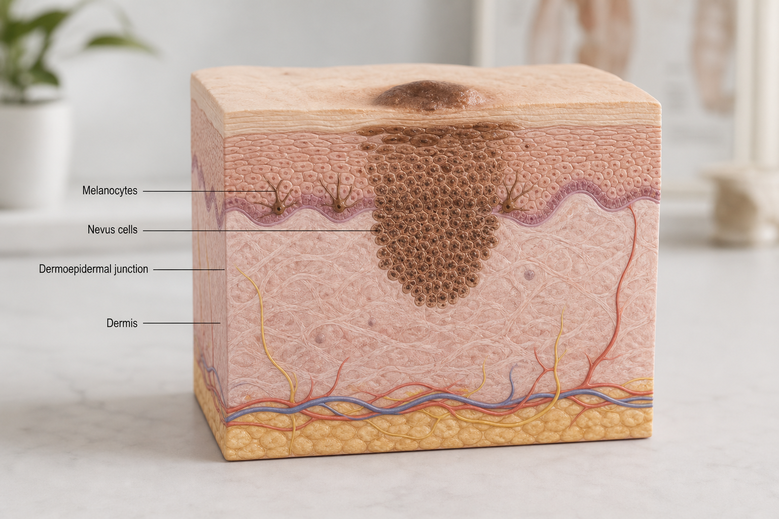

Noticing pigment returning to a site where a mole was removed can be unsettling, but it is a recognised clinical phenomenon with a clear explanation. Moles can grow back after removal, and in most cases the regrowth is benign — the result of residual mole cells left behind rather than anything more serious. Understanding why regrowth happens, what it looks like, and when it warrants further assessment can help you respond calmly and appropriately.
Key Takeaways
- Mole regrowth is most commonly caused by incomplete removal, where residual mole cells remain in the deeper layers of the skin and repopulate the scar.
- Shave excision carries the highest regrowth risk — estimated at roughly 11–30% — while full-thickness surgical excision with clear margins has the lowest.
- Returning pigment is usually benign, but pigment spreading beyond the scar edge requires prompt dermatological assessment to rule out melanoma.
- The clinical term for benign mole regrowth is recurrent nevus, sometimes called pseudomelanoma because it can mimic melanoma under dermoscopy.
- Most regrowth appears within three months of the procedure; any new pigment appearing after six months or showing ABCDE warning signs should be reviewed.
Can Moles Grow Back After Removal?
Yes, moles can grow back after removal, and this is more common than many patients expect. The likelihood depends primarily on the removal method used and whether the procedure achieved complete removal of all nevus cells. A mole is not simply a surface mark — it is a cluster of melanocytes and nevus cells that can extend into the deeper layers of the dermis. If any of those cells remain after a procedure, they retain the capacity to proliferate and produce pigment again, causing visible repigmentation at the treated site.
Regrowth does not automatically indicate a problem with the original procedure or a sign of cancer. It is a well-documented biological response to incomplete removal, and dermatologists have a specific clinical term for it: recurrent nevus. What matters most is how the returning pigment behaves, where it appears relative to the scar, and whether it displays any features that warrant further investigation.
Why Does Mole Regrowth Happen? The Mechanism Explained
Every mole contains melanocytes — the pigment-producing cells of the skin — along with specialised nevus cells that cluster at the dermoepidermal junction and within the dermis. When a mole is removed, the goal is to eliminate all of these cells. If even a small population of nevus cells is left behind, they can survive within the healing scar tissue and gradually multiply, eventually producing enough melanin to become visible at the skin surface.
This process is sometimes described using an iceberg analogy: the visible pigmented surface of a mole represents only the portion above the waterline, while the deeper cellular component extends further into the dermis than it appears. Removal techniques that address only the surface portion — such as shave excision — are more likely to leave mole cells behind in the deeper dermis, making regrowth more probable.
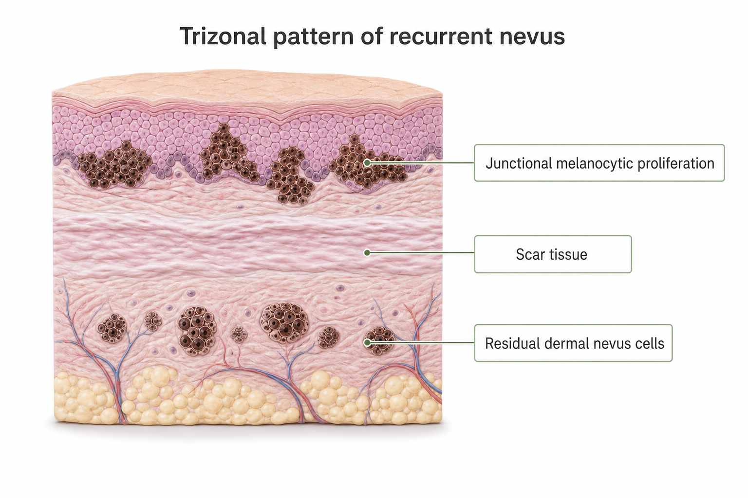
The resulting clinical picture is known as the recurrent nevus phenomenon. Histologically, it displays a characteristic trizonal pattern: a layer of junctional melanocytic proliferation at the top, a band of scar tissue in the middle, and residual dermal nevus cells below. This pattern is important because it helps pathologists distinguish benign regrowth from melanoma when a re-biopsy is performed.
Which Removal Methods Carry the Highest Regrowth Risk?
The removal technique is the single most important factor determining whether a mole grows back. Different methods reach different depths of the skin, and this directly affects how completely the nevus cells are eliminated.
| Removal Method | Depth Reached | Estimated Regrowth Risk | Histopathology Available? |
|---|---|---|---|
| Shave excision / shave biopsy | Superficial dermis | Approximately 11–30% | Yes (partial sample) |
| Full-thickness surgical excision | Full dermis to subcutis | Very low when clear margins achieved | Yes (complete sample) |
| Laser removal | Variable; typically superficial | Moderate to high; tissue destroyed, not sampled | No |
| Cryotherapy | Superficial to mid-dermis | Moderate; depth control limited | No |
| Radiofrequency (RF) surgery | Superficial to mid-dermis | Moderate; depends on technique | No (tissue vaporised) |
| At-home removal products | Unpredictable | High; no depth control | No |
Shave Excision and Regrowth
Shave excision — also called shave removal or shave biopsy — is one of the most commonly performed mole removal procedures. A blade is used to shave the mole flush with or just below the skin surface, removing the raised portion without cutting deeply into the dermis. This approach produces a good cosmetic result and allows the removed tissue to be sent for histopathology, but it is inherently more likely to leave residual mole cells in the deeper dermis. Published regrowth rates for shave excision range from approximately 11% to 30%, with the majority of recurrences appearing within three months of the procedure.
Full-Thickness Surgical Excision
Full-thickness excision involves cutting the mole out completely, including a margin of surrounding normal tissue, and closing the wound with sutures. When the excised tissue is examined and confirmed to have clear margins — meaning no nevus cells are detected at the edges of the removed specimen — the risk of regrowth is very low. This method is the standard approach for moles with atypical features or any clinical concern about malignancy, and it provides a complete histopathology sample for assessment.
Laser, Cryotherapy, and RF Removal
Laser removal, cryotherapy, and radiofrequency surgery destroy mole tissue rather than excising it. Because no tissue sample is collected, histopathology is not possible, which means any atypical or cancerous cells cannot be identified after the procedure. These methods also offer less precise depth control than surgical excision, making incomplete removal more likely for deeper moles. For these reasons, most dermatologists do not recommend laser or cryotherapy for moles that have not been assessed and confirmed as benign.
At-Home Removal Risks
Over-the-counter creams, acids, and DIY removal kits marketed for mole removal carry significant risks. They offer no depth control, no histopathology, and no way to confirm whether the mole has been fully or safely removed. Beyond a high regrowth risk, at-home removal can cause scarring, infection, and — critically — can obscure a mole that may have required proper clinical assessment. Dermatologists consistently advise against self-removal of moles.
Does a Mole Growing Back Mean Cancer?
In the vast majority of cases, mole regrowth after removal is benign. The returning pigment represents residual nevus cells repopulating the scar — not malignant transformation. However, the appearance of a recurrent nevus can be genuinely alarming, and for good reason: under dermoscopy, benign regrowth can display irregular pigment patterns that closely resemble melanoma. This is why the condition is sometimes referred to clinically as pseudomelanoma.
The key distinction that dermatologists use is the relationship between the returning pigment and the scar boundary. Pigment confined within the scar is generally consistent with benign recurrent nevus — the cells are simply repopulating the area where the original mole sat. Pigment that spreads beyond the scar edge into surrounding normal skin is a more concerning finding that warrants urgent re-evaluation, as this pattern is less typical of simple regrowth and more consistent with a process that requires histopathological assessment.
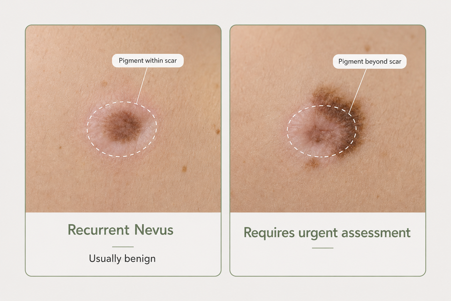
It is also important to apply the ABCDE rule to any returning pigment: Asymmetry, irregular Borders, multiple Colours, a Diameter greater than 6mm, and Evolution (change over time). Any of these features in a regrowing mole should prompt a dermatology appointment without delay.
What Does Mole Regrowth Look Like? Scar vs. Regrowth
Distinguishing normal scar healing from genuine mole regrowth is not always straightforward, particularly in the first few weeks after a procedure. During healing, the skin may appear pink, slightly raised, or discoloured — none of which indicates regrowth. True mole regrowth involves the reappearance of brown or dark pigment within the healed scar, typically becoming visible between one and three months after the procedure.
Hair Regrowth vs. Mole Regrowth
Patients sometimes mistake hair regrowth for mole regrowth, particularly when the original mole was raised or hairy. Hair follicles can survive a shave excision and produce new hairs within the scar area. This is not the same as mole regrowth — the hair follicle itself is not a nevus cell, and its reappearance does not indicate that the mole is returning. If you notice dark hairs growing from a healed scar but no flat pigmented area developing, this is likely hair follicle activity rather than nevus cell repopulation.
Post-Inflammatory Pigmentation
Post-inflammatory pigmentation is another phenomenon that can be confused with mole regrowth. After any skin injury or procedure, melanocytes in the surrounding skin may produce excess pigment as part of the healing response, creating a flat, diffuse discolouration around the scar. This type of pigmentation tends to be more uniform in colour, lacks the structured pattern of a recurrent nevus, and often fades over several months. It is more common in people with darker skin types. A dermatologist can usually distinguish post-inflammatory pigmentation from true regrowth during a clinical or dermoscopic assessment.
Understanding Pseudomelanoma: Why Regrowth Can Look Alarming
The term pseudomelanoma — formally known as recurrent melanocytic nevus — describes the histological and dermoscopic appearance of a benign regrowing mole that mimics melanoma. Under dermoscopy, a recurrent nevus can display atypical pigment networks, irregular streaks, and asymmetric colouring that would raise concern if seen in a primary lesion. This is why even experienced dermatologists may order a re-biopsy when they encounter regrowth, particularly if the original histopathology report is unavailable.
The trizonal histologic pattern — junctional proliferation above the scar, scar tissue in the middle, residual dermal nevus cells below — is the pathological hallmark that allows a pathologist to confirm the diagnosis of recurrent nevus rather than melanoma. This is one reason why retaining the original histopathology report from your mole removal is genuinely useful: it gives any future dermatologist the baseline information needed to interpret regrowth accurately and avoid unnecessary procedures.
How Long After Removal Can a Mole Grow Back?
Most mole regrowth occurs within the first three months after removal. One published figure suggests that approximately 94% of recurrences become visible within this window. Regrowth appearing between three and six months is less common but still possible. New pigmentation appearing more than six months after a procedure — particularly if it was not present during the initial healing phase — is less likely to represent simple regrowth and should be assessed by a dermatologist, as it may reflect a different process.
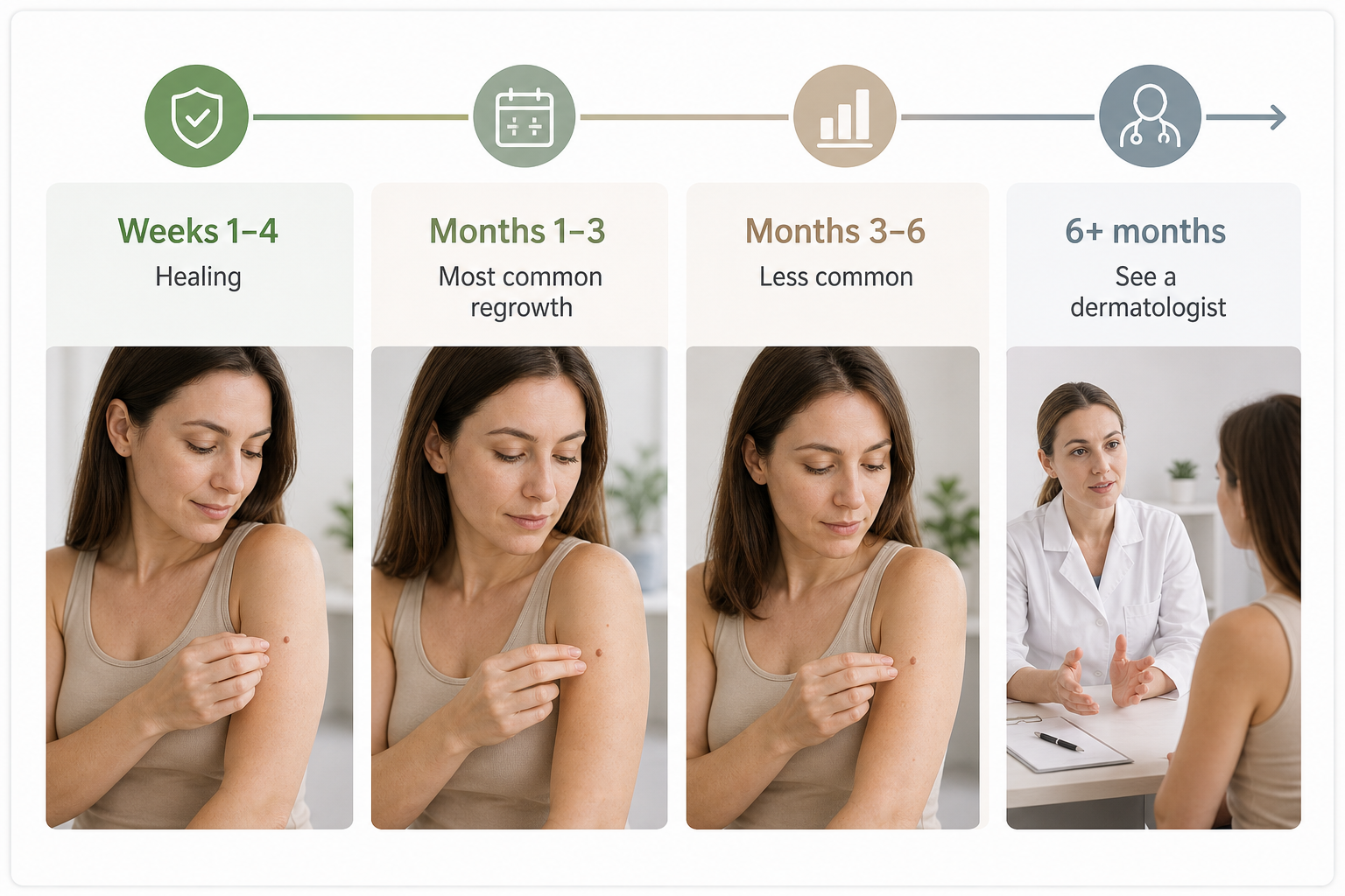
- Weeks 1–4: Healing phase — redness, scabbing, and early scar formation are normal. No pigment should be interpreted as regrowth at this stage.
- Months 1–3: The most common window for true mole regrowth to become visible. Pigment within the scar boundary is usually benign.
- Months 3–6: Less common regrowth window. Any new pigment should be monitored and assessed if it changes.
- Beyond 6 months: New pigmentation at a previously clear removal site warrants dermatological review, particularly if it shows ABCDE features or extends beyond the original scar.
What to Do If Your Mole Grows Back
If you notice pigment returning to a mole removal site, the appropriate first step is to arrange a review with a dermatologist rather than attempting to monitor it independently or seek further cosmetic removal. A dermatologist will assess the returning pigment using dermoscopy — a non-invasive technique that uses magnification and polarised light to examine pigment patterns in detail — and compare the findings against your original histopathology report if one is available.
If the dermoscopic appearance is reassuring and the pigment is confined within the scar, the dermatologist may recommend monitoring with periodic skin checks or mole mapping using digital imaging. If the appearance is atypical, or if the original mole was not sent for histopathology, a re-biopsy will typically be recommended. This involves removing a tissue sample from the regrowing area and sending it for histopathological analysis to confirm whether the cells are benign or require further management.
What Re-Excision Involves
If re-excision is recommended, the procedure generally involves removing the regrowing area along with a margin of surrounding tissue, using full-thickness surgical excision to ensure clear margins. The excised tissue is sent for histopathology, and the results are reviewed against the original pathology report. If the original mole was benign and the regrowth confirms a recurrent nevus with clear margins on re-excision, no further treatment is usually required. If atypical cells are identified, the dermatologist will advise on appropriate next steps, which may include wider excision.
Risk Factors That Make Regrowth More Likely
Certain characteristics of the mole and the patient can increase the probability of regrowth after removal. Being aware of these factors can help set realistic expectations before a procedure.
- Removal method: Shave excision carries a significantly higher regrowth risk than full-thickness excision with clear margins.
- Mole depth: Deeper moles — particularly compound nevi and dermal nevi — extend further into the dermis and are more likely to have residual cells after superficial removal.
- Raised or hairy moles: These tend to have a larger dermal component, increasing the chance of incomplete removal.
- Multi-coloured moles: Moles with varied pigmentation may have a more complex cellular architecture that is harder to remove completely.
- Younger patients: Younger skin has a more active healing response, and residual nevus cells may proliferate more readily.
- Darker skin types: Post-inflammatory pigmentation is more common and can complicate the clinical picture.
- Facial location: Facial skin has a rich blood supply and relatively thin dermis, which may support more active repigmentation after incomplete removal.
- Congenital moles: Moles present from birth often have deeper and more extensive cellular involvement than acquired moles.
Skin Cancer Monitoring After Mole Removal
Mole removal — whether for cosmetic reasons or due to clinical concern — does not eliminate the need for ongoing skin cancer monitoring. People who have had moles removed, particularly those with a history of atypical or dysplastic nevi, should continue with regular skin checks. Annual full-body skin examinations by a dermatologist are generally recommended for anyone with a personal or family history of melanoma, multiple atypical moles, or significant UV exposure history.
Mole mapping using digital dermoscopy allows a dermatologist to photograph and record the appearance of multiple moles over time, making it easier to detect subtle changes that might not be apparent during a single examination. This is particularly useful for patients with many moles or a history of atypical features, and it provides a documented baseline against which any future changes — including regrowth at a removal site — can be compared.
Frequently Asked Questions
Is it normal for a mole to grow back after shave removal?
Yes, regrowth after shave excision is a recognised and relatively common outcome, with published rates ranging from approximately 11% to 30%. Shave removal addresses the surface portion of the mole but may leave residual mole cells in the deeper dermis. If pigment returns within the scar boundary and does not display atypical features, this is usually benign. A dermatologist can confirm this with dermoscopy and, if needed, a re-biopsy.
Can laser mole removal cause regrowth?
Yes. Laser removal destroys mole tissue rather than excising it, which means depth control is limited and no tissue sample is available for histopathology. If laser energy does not reach all the nevus cells, regrowth can occur. Because laser removal does not allow pathological assessment, most dermatologists advise against it for moles that have not been clinically confirmed as benign.
Does removing a mole spread cancer?
There is no reliable clinical evidence that removing a benign mole causes cancer or spreads cancerous cells. This is a common misconception. However, if a mole is malignant and is removed incompletely — without clear margins — the remaining cells can continue to grow. This is why proper histopathological assessment and clear margins are important, particularly for any mole with atypical features.
How can I tell if regrowth is benign or concerning?
The most important clinical distinction is whether the returning pigment is confined within the mole removal scar or extends beyond it. Pigment confined within the scar is generally consistent with benign recurrent nevus. Pigment spreading beyond the scar edge, or pigment that is asymmetric, has irregular borders, multiple colours, or is growing rapidly, should be assessed by a dermatologist promptly. Do not attempt to self-diagnose regrowth.
Will a regrowing mole keep growing indefinitely?
Not necessarily. Some recurrent nevi stabilise and do not continue to enlarge significantly. However, because regrowth can be difficult to distinguish from melanoma without dermoscopy or histopathology, any regrowing mole should be assessed by a dermatologist rather than simply observed at home. If re-excision with clear margins is performed, the risk of further regrowth is substantially reduced.
Can I prevent mole regrowth?
The most effective way to minimise regrowth risk is to choose a removal method that achieves complete removal — ideally full-thickness surgical excision with histopathological confirmation of clear margins. Protecting the treated area from UV exposure during healing may reduce post-inflammatory pigmentation, though it does not prevent true nevus cell regrowth. Avoiding at-home removal products and ensuring the procedure is performed by a qualified practitioner also reduces risk.
When Mole Regrowth Appears: What Your Next Step Should Be
Seeing pigment return to a mole removal site is understandably worrying, but the clinical reality is that most regrowth represents benign recurrent nevus — residual mole cells repopulating a healing scar — rather than anything malignant. The key is not to ignore it or attempt further self-treatment, but to have it properly assessed. A dermatologist can use dermoscopy to evaluate the returning pigment, review your original histopathology report, and determine whether monitoring, re-biopsy, or re-excision is appropriate.
If you are planning a mole removal and want to minimise regrowth risk, discussing the available methods with a qualified practitioner before the procedure is worthwhile. Full-thickness surgical excision with clear margins offers the most reliable outcome for complete removal, particularly for raised, deep, or multi-coloured moles. Whatever method is used, retaining a copy of your histopathology report gives any future clinician the baseline information needed to interpret changes accurately. Ongoing skin cancer monitoring — including annual skin checks and mole mapping where appropriate — remains important regardless of which moles have been removed.
